Information engineer Bruno Sanches carefully places an orange, conical apparatus of 15 centimeters (cm) in diameter, which looks similar to the cup of a brassiere, onto the table at the University of São Paulo Polytechnic School’s Integrated Systems Laboratory (POLI-USP). And not without good reason: this is the prototype of a new generation of examination devices that adjust around the breast and capture images at different angles in the search for tumors that need to be removed. The Brazilian National Cancer Institute (INCA) estimated 73,600 new cases of breast cancer per year in the country between 2023 and 2025. It is the most common tumor among women across all regions of Brazil, with the highest rates in the country’s South and Southeast.
The key characteristic of the new imaging system, a joint project between POLI-USP and the Electrical Engineering Department at the Federal Institute of São Paulo (IFSP), is its microwave function, a new technological approach also studied at universities in the US, Canada, UK, Sweden, Italy, Japan, and Australia. Mammography devices currently used to detect breast tumors operate with X-ray, a type of ionizing radiation that can present risks to health.
Although well tolerated by most women, the traditional scan can cause discomfort and pain due to compression of the equipment’s plates on the breasts. The new device designed by the São Paulo researchers molds to the breast shape and avoids the painful process. In 2023, 4.4 million mammographies were conducted by the Brazilian Public Health System (SUS).
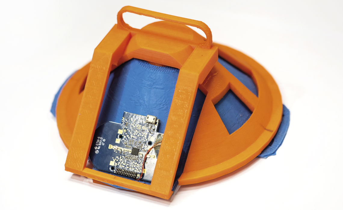
Léo Ramos Chaves / Revista Pesquisa FAPESPPrototype of microwave imaging system designed by USP and IFSPLéo Ramos Chaves / Revista Pesquisa FAPESP
“We want to offer a complementary alternative to the mammography,” says Sanches. The engineer believes the device could be especially useful among women with denser breasts, for which the mammography is less sensitive. Density, which is not related to size, is the proportion of fibrous and glandular tissue in relation to adipose (fat) tissue.
The standard of the American Radiology College (ACR) for imaging scans classifies the breasts into four densities: predominantly fatty and less dense, easier to scan by mammography; those with sparse areas of fibroglandular tissue; heterogeneously dense, which may hamper the detection of small nodules or tumors; and extremely dense, making the identification of lesions on the mammograph even more difficult. The proposed system may be advantageous for dense breasts, as the electromagnetic properties of the tissues involved are discrepant and can be differentiated.
“The denser the breast, the whiter it appears on the mammograph, making it difficult to identify tumors, which are also white,” explains radiologist physician Almir Bitencourt, of São Paulo’s A.C.Camargo Cancer Center, a leading Brazilian facility for research, diagnosis, and treatment in the area. “In these situations, complementary exams such as ultrasound or magnetic resonance imaging (MRI) are normally recommended.”
Radio waves
The prototype developed by USP and IFSP uses an electronic device, known as a microwave transceiver, that transmits and receives signals through embedded antennae. The transceiver emits ultrabroadband radio waves at a central frequency of 6.4 gigahertz (GHz), which pass through the mammarian tissue and return to the device if they find denser internal structures, such as possible tumors. The reflected signals are directed to an image processing unit, which uses an algorithm to generate a detailed map of the region (see infographic).
The system currently generates reconstructed two-dimensional images of the breast, but the hardware structure enables adjustments to the vertical position of the platform, allowing the antenna locations to be varied. The equipment can then scan different sections of the breast, producing 3D images.
In tests on an artificial model, known as the phantom, with materials aimed at replicating the electrical properties of the mammary tissues, researchers found that the device can detect tumors of 1 cm in diameter and at 3 cm depth, as detailed in a January 2023 article published in the journal Biomedical Signal Processing and Control.
The internal structure of the phantom was designed to simulate breast anatomy, with 0.2 cm of skin, 6 cm of glandular tissue, and 8.6 cm of fat tissue. Breast cancer is classified into four clinical stages according to its extent and severity. Tumors up to 2 cm in diameter not reaching the lymph nodes are in the initial stage and are less serious.
Recommended for the diagnosis of suspicious alterations at any age in both men and women, the X-ray mammogram can identify lesions smaller than 1 cm, along with the initial signs of breast cancer, known as microcalcifications. “Currently, no other method identifies microcalcifications with the same accuracy,” says Bitencourt, of the A.C.Camargo Center.
The microwave exam, although less accurate in its current version, may put a stop to the ionizing radiation of traditional devices. “X-ray mammography needs shielded spaces, while the microwave-based technology does not emit ionizing radiation, making it safer and more accessible,” says electrical engineer Fatima Salete Correra, also of POLI-USP, who did not participate in the research. Low-cost, portable devices may bring benefits, particularly in regions where access to breast scans is more challenging.
The team’s previous experience with the miniaturized integrated circuit known by the abbreviation SAMPA (serialized analog-digital multi-purpose Asic), developed with the support of FAPESP (see Pesquisa FAPESP issue nº 253), helped in the development of components for integration into the portable prototype. “Knowledge about how to design and build highly sensitive amplifiers, analog-digital converters, and signal processors, was essential to our development of chips for use in healthcare,” explains Sanches, of the Polytechnic. Created by researchers from USP, UNICAMP, and the Aeronautics Technology Institute (ITA), the SAMPA Chip has been in use since 2020 at one of the four particle detectors of the Large Hadron Collider (LHC), operated by the European Organization for Nuclear Research (CERN) on the French-Swiss border.
The next step for the São Paulo team will be to test the performance of the prototype on phantoms with different breast sizes and tumor types. Companies are yet to show an interest in collaborating on the project. In other countries, devices of this type are already in more advanced stages of development. In a review article published in December 2024 in IEEE Access, the researchers from USP and IFSP compared the performance of 12 prototypes, and saw how they present different levels of sensitivity (ability to correctly identify people with a tumor) and specificity (ability to present a negative result with no tumor present).
Some demonstrated high levels of precision, such as the device at McMaster University, Canada, which was found to identify tumors of 2.4 mm, though the exam takes five hours. One of the most advanced devices, created at the University of Bristol in England, is known as Maria (multistatic array processing for radio-wave image acquisition). In a clinical trial involving 389 women with an average age of 47, the sixth version of the device correctly identified 47% of malign lesions, falling well short of the percentage for conventional mammography, whose correct identification rate was 92%, as detailed in the British Journal of Radiology in a January 2024 article.
Although the scan was well evaluated by the women, the authors of the study, led by radiologist Richard Sidebottom of the Royal Marsden NHS Foundation Trust, concluded that diagnosis by microwave cannot yet be considered fully effective. Of the total participants, 94% preferred this procedure to the traditional scan, primarily due to there being no breast compression or ionizing radiation. Nine out of ten women reported better comfort during diagnosis.
The story above was published with the title “Microwaves to hunt tumors” in issue in issue 350 of april/2025.
Scientific articles
DE JESUS ARAGÃO, A. et al. Low-cost device for breast cancer screening: A dry setup IR-UWB proposal. Biomedical Signal Processing and Control. Vol. 79, 104078. Jan. 2023.
DE JESUS ARAGÃO, A. et al. A review on microwave Imaging Systems for Breast Cancer Detection. IEEE Access. Vol. 12, pp. 190611–28. Dec. 2024.
SIDEBOTTOM, R. et al. Results for the London investigation into dielectric scanning of lesions study of the MARIA® M6 breast imaging system. British Journal of Radiology. Vol. 97, no. 1155, pp. 549–52. Jan. 24, 2024.
Republish