
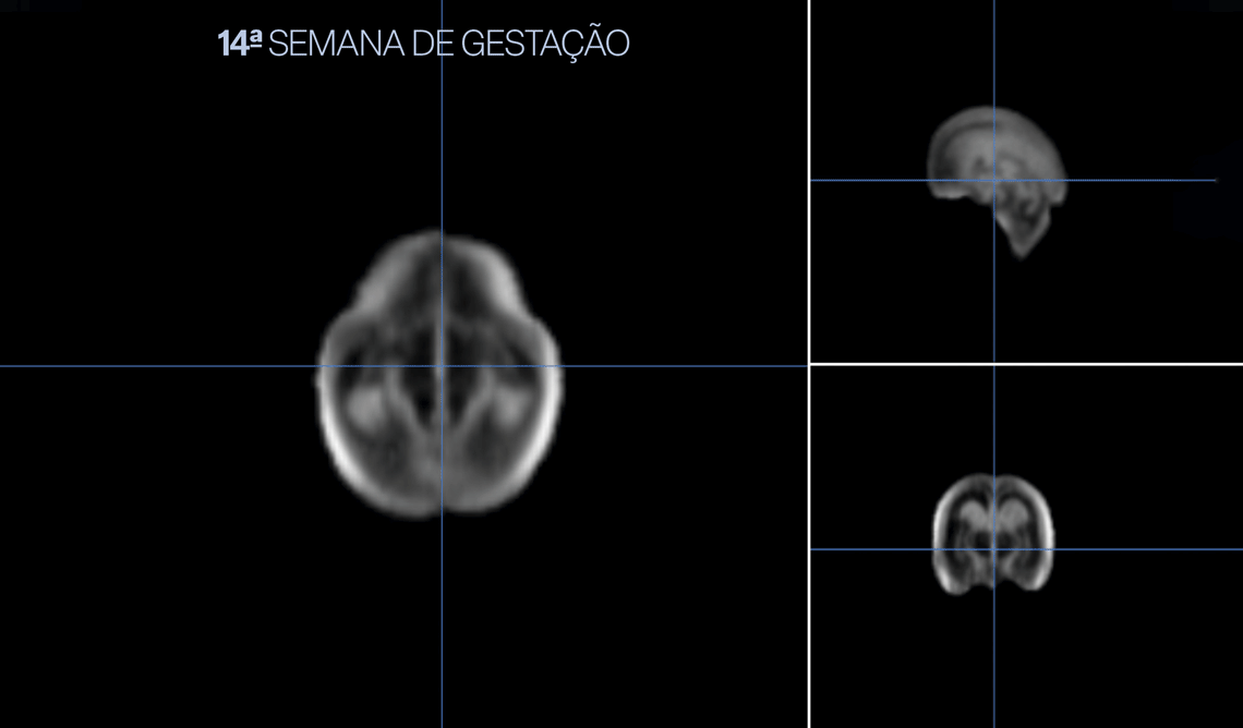
High-resolution ultrasound images of 899 babies have allowed an international group of researchers to accurately map the brain and encephalon development of healthy fetuses in the womb. Organized in an online atlas, this study’s findings should serve as a reference for expected and desired brain development during pregnancy. It should also help doctors detect, at an earlier stage, any damage or problems with neurological maturation that could lead to future impairment. The study was supervised by obstetrician José Villar and human reproduction specialist Stephen Kennedy, both from Oxford University, in the UK, with support from Brazilian researchers, and was published in the October edition of Nature.
To arrive at the parameters of what appears to be optimal human brain development in the womb, almost 200 researchers tracked 899 pregnancies across eight countries — including Brazil — in the Americas, Europe, Asia, and Africa from 2009 to 2016. The specialists performed 3D ultrasounds of the fetuses periodically, from 14 weeks gestation until shortly before delivery, ensuring that the same equipment and standardized procedures were always used, enabling them to reconstruct brain evolution week by week. All the mothers were in good health and had the highest levels of education within their communities. None of them smoked or had any illnesses that could compromise the baby’s development.
The reason this group was selected is because women with more schooling tend to take better care of their health and are at a lower risk of experiencing problems during pregnancy. By solely monitoring fetuses carried under the best conditions, we can determine supposedly ideal development of brain structures and other encephalon components.
“Our atlas fills a six-week knowledge gap in the understanding of how the brain matures in early fetal life,” states Ana Namburete, a researcher at Oxford University and lead author of the study, in a press release. “We also revealed significant asymmetries in brain maturation, such as in the region associated with language development, which peaked between 20 and 26 weeks gestation and persisted without differences between sexes,” she stated.
Measuring a fetus’s brain structures is no simple task. From 14 to 31 weeks gestation, the period detailed in the current atlas, the brain’s volume grows by 13 times, from 24 cubic millimeters (mm3) to 318 mm3. During this phase, cells multiply rapidly and establish interconnections. Cell groups migrate to specific regions, making up the control nuclei for different functions. “During this period, neuronal proliferation defects can occur, leading to clinical consequences,” explains neuroscientist Andrea Jackowski, of the Federal University of São Paulo (UNIFESP), who studies brain development but was not involved in this study. “There are moments in which the brain is more vulnerable to environmental factors and any form of aggression that can have repercussions for the child later. But this stage can also serve as a window of opportunity to intervene and fix problems,” she states.
Asymmetries
Another factor that makes it difficult to obtain measurements is that, during this period, the right and left hemispheres are starting to become asymmetrical, with some regions gaining more volume in one hemisphere than the other. This means that the brain assumes the shape it will have for the rest of its life while in the womb, even if it is not entirely mature and undergoes changes later. “It is a fundamental phase of maturation in which, after the initial formation, the brain structures experience regional specialization and development processes. It lays the foundation for babies to be ready for their first acts of cognitive development when they are born,” explains psychiatrist Pedro Pan, also from UNIFESP but who was not involved in the study described in Nature.
Added to these changes is the fact that the baby moves around a lot in the womb, complicating image retrieval. The current study has improved imaging by using artificial intelligence. “The authors had to use machine learning techniques and deep neural networks, in addition to complex image processing methods, to compile the atlas,” explains statistician and neuroscientist João Ricardo Sato, of the Federal University of ABC (UFABC), who reviewed the study at the request of Pesquisa FAPESP.
Several groups had already recorded brain development in human fetuses, but they covered a later period (from 19 weeks gestation) and had fewer participants (from 12 to 197) — almost always seen at a single clinic and never followed up with after birth.
The study published in Nature included pregnant women from different ethnic groups and followed up with the children until their second year of life. It is part of the International Fetal and Newborn Growth Consortium for the 21st Century (INTERGROWTH-21st), which followed almost 20,500 children from pregnancy to their first years of life and helped produce growth curves from intrauterine life to 2 years (see Pesquisa FAPESP issue nº 225).
“This is the largest study, to our knowledge, describing the early normative development of the human brain,” wrote the authors of the Nature article. “Our reconstructions of the fetal brain provide the first in vivo representation of the entire second trimester [of pregnancy],” they affirmed.
“The data establishes a normal pattern of fetal brain growth, which is important for diagnosing neurological problems at this stage of life,” explains pediatrician and epidemiologist Fernando Barros, of the Federal University of Pelotas (UFPel), coauthor of the study published in Nature, and coordinator of Brazil’s participation in INTERGROWTH-21st, which provided 86 fetus images for the study. “The atlas can become a valuable tool for healthcare professionals, aiding in clinical decision-making and prenatal care, as well as a useful tool for education,” he states.
“Regular ultrasound exams allow problems to be identified as of 22 weeks gestation. With the development pattern presented in the atlas, we may be able to detect changes earlier,” concludes obstetrician Denise Lapa, a fetal surgery specialist at Hospital Israelita Albert Einstein, in São Paulo.
Scientific article
NAMBURETE, A. I. L. et al. Normative spatiotemporal fetal brain maturation with satisfactory development at 2 years. Nature. Oct. 25, 2023.