Eduardo GorabTriple helix (pink) identified in the chromosome of a fly: gene regulationEduardo Gorab
Four years ago, researcher Eduardo Gorab, of the University of São Paulo Biosciences Center (IB/USP), developed a method that uses a known antibody to recognize a rare type of structure found in the genetic material of flies of the species Rhynchosciara americana and Drosophila melanogaster: DNA molecules made up of three twisted strands of nitrogenous bases instead of the traditional double helix, which is the standard shape of deoxyribonucleic acid, or DNA. This unusual triple helix was observed in the heterochromatin, a chromosomal region where the DNA is compacted with proteins and RNA (ribonucleic acid). When Gorab identified the triple helix in this region, he suspected that it might be associated with the gene inactivation process, which is common in the heterochromatin. However, a study published on January 27 of this year in the science journal Nature Structural & Molecular Biology by this Brazilian researcher and colleagues from Europe and Japan suggests that these triple helices may play other roles in the cell nucleus.
Using the molecular tool designed by Gorab, the international group of researchers found triple helices formed of chains of bases whose structure differed slightly from that identified in the genetic material of the flies; these were located in the chromatin of mouse embryos. Instead of three twisted strands of DNA, the animal cells displayed two DNA strands interwoven with one RNA strand. These triple helices were identified at a very specific, early stage in the process of embryo development, when the latter consisted of only two or eight cells. During this stage of embryogenesis, the presence of the triple helices seemed to increase the expression of certain genes that are important in this phase in the process. In more advanced embryo stages, when this set of genes was no longer activated, the RNA strand was also no longer observed to be bonded with the two DNA strands. “We also saw that when we stimulated the production of the triple helix in vivo, the expression of these genes increased,” states Gorab. “These findings do not constitute direct, decisive proof that this happens, but they reinforce the correlation.”
According to one of the study’s authors, researcher Maria-Elena Torres-Padilla, with the Institute of Genetics and Molecular and Cellular Biology (IGBMC) in Strasbourg, France, the possible regulatory role of the triple helix appears at this stage in embryo development, based on atypical chromatin architecture. By definition, chromatin displays two different forms: an active one, known as euchromatin, in which DNA is accessible and can be expressed by regulatory proteins, and an inactive one – the heterochromatin mentioned earlier – in which genetic material is compacted and cannot be used.
 In the research on rodent embryo cells, chromatin was at an atypical stage, intermediary between its two forms, where it could be accessed and regulated by the triple helix. “We were looking for an RNA-based regulatory mechanism that would have an impact on the structure, or ‘conformation’ of the chromatin,” explains Torres-Padilla. “Since RNA, together with DNA, forms a triple helix, it was a good candidate for playing this role.”
Although the phenomenon of DNA molecule formation entailing more than two strands of nucleic acid has been under study since the 1950s, it has only been in the last 10 to 15 years that biochemists began to gain a better understanding of the mechanisms that can lead to the appearance of this less conventional kind of genetic material. “Triple helices tend to form in genomic regions where there are frequent repeated sequences of a base, although there are other possibilities as well,” says Gorab. In other words, stretches of DNA rich in sequences with a single nucleotide, like TTTTT (thymine) or AAAAA (adenine), are candidates for harboring helices with more than two strands. Since about half of the mammalian genome is composed of repetitive sequences, formed of transposable elements (transposons and retrotransposons) that can change position or copy themselves along the genome, the presence of this type of structure is probably not all that rare.
Quadruple helix DNA
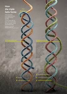
Three-stranded DNA can form in more than one way. In Gorab’s research with flies, the triple helix originated from the separation of the two strands that make up the standard DNA molecule, starting at a certain point along the sequence. On one piece of DNA, the two chains of nitrogenous bases stay paired while the other piece presents loose strands. One of these loose strands bends back and wraps itself around the stretch of DNA where the two chains are still paired. This yields a triple-stranded DNA molecule, all strands of which originated from a single molecule. This is intramolecular triple-stranded DNA.
There is also an intermolecular DNA triplex, which occurs when one of the strands is donated by a second DNA molecule. In this case, two chains of the triple helix come from a conventional DNA molecule while another strand detaches itself from another DNA molecule. In the research with mice in which Torres-Padilla used the antibody created by Gorab, the third DNA strand was donated by an RNA molecule, which usually displays only one chain of bases. “Our methodology can identify a number of forms of triple-helical nucleic acids,” says the USP researcher.

Gabriel BitarQuadruple DNA structure: a possible link to cancerGabriel Bitar
There is no evidence that the formation of genetic structures about which scientists know little as yet – like the triple helix – is necessarily associated with the development of disease. Theoretically, elements that act to regulate gene activity can produce positive as well as negative effects. A study published this January in the journal Nature Chemistry identified quadruple helix DNA – another unusual conformation for this molecule – in human cancer cells. The discovery may prove useful in the effort to understand the process by which tumors appear and perhaps even in the development of new treatment approaches.
With four interlaced strands, this type of DNA forms along genomic stretches rich in guanine, a nitrogenous base represented by the letter ‘G’ – thus the names G-quadruplex or G-quartet. “Research shows that quadruplexes occur more often in the genes of cells that are dividing rapidly, like cancer cells,” the lead author of the study, Shankar Balasubramanian, of the University of Cambridge, stated at the time of publication. “For us, it strongly supports a new paradigm to be investigated: using these four-stranded structures as targets for personalized treatments in the future.”
Project
Molecular aspects of heterochromatin in species from the family Sciaridae (Diptera: Nematocera) (2008/50653-2); Grant Mechanism Regular Line of Research Project Award; Coordinator Eduardo Gorab – IB/USP; Investment R$165,485.11(FAPESP).
Scientific article
FADLOUN, A. et al. Chromatin signatures and retrotransposon profiling in mouse embryos reveal regulation of LINE-1 by RNA. Nature Structural & Molecular Biology. 27 Jan. 2013.