 Léo RAmosEarly on the afternoon of March 13, 2015, during a break in an international scientific meeting on brain mapping, a new research facility at the University of São Paulo School of Medicine (FMUSP) will be inaugurated. Called an Imaging Platform in the Autopsy Room (PISA), the laboratory was built underground and occupies an area of 500 square meters. Land adjacent to FMUSP’s headquarters was excavated to accommodate the facility, which houses the Magnetom 7T MRI, the first magnetic resonance imaging equipment in Latin America capable of scanning the entire body. It has an extremely powerful magnetic field of 7 Tesla. The equipment will be used primarily to study cadavers arriving at the Death Certification Service of the City of São Paulo (SVOC), which is maintained by USP. Each year it performs around 14,000 autopsies on natural deaths (violent deaths are handled by the Medical Examiner’s Office). One of the objectives of the research is to develop diagnostic imaging techniques to help identify the cause of death in a less invasive way than conventional autopsies. Studies of the dead promise to help the living through advanced techniques in the diagnosis and understanding of diseases. “In diagnostics, we should have immediate feedback,” says Dr. Paulo Hilário Saldiva, a professor of pathology at FMUSP and the project’s coordinator.
Saldiva is referring to diseases affecting organs that are difficult to study while the patient is alive, since removal of tissue is risky. “There has never been as much chemotherapy as today, and some patients end up having heart problems because these drugs are toxic to the heart. One idea is to perform a minimally invasive autopsy on people who have succumbed to such heart problems and obtain samples of their heart tissue. This work can be done quickly, in 15 or 20 minutes, causing only a slight delay in the release of the body to the family of the deceased.”

ERWIN HAHN INSTITUTE FOR MR 2 MEDICAL UNIVERSITY, VIENA 3 PETER MORRIS, NOTTINGHAM UNIVERSITY7 Tesla magnetic resonance images ERWIN HAHN INSTITUTE FOR MR 2 MEDICAL UNIVERSITY, VIENA 3 PETER MORRIS, NOTTINGHAM UNIVERSITY
Among the possibilities that are beginning to appear, Saldiva also cites research on what are known as solitary pulmonary nodules that appear separately in diagnostic tests, but about which little is known, since in most cases no biopsy is indicated. Patients have to undergo control tests. It is possible to take samples of these nodules in minimally invasive autopsies and generate information about their characteristics. The director of the SVOC, Dr. Carlos Augusto Pasqualucci, who is a professor with FMUSP’s Department of Pathology, highlights the project’s multiple approaches. “We want to improve our techniques for investigating the causes of natural death and develop more sensitive tests for disease diagnosis,” he says. “The idea is to use magnetic resonance images so that radiologists can better understand the nature of changes in organs and tissues and make better diagnoses.”
“We will work with families on another idea of giving, providing us with knowledge, demonstrating the importance of studying cadavers to advance the understanding of diseases,” says Saldiva. “The 7 Tesla equipment is being used in other parts of the world, but none operates in an environment as fertile for research as ours.” The director of USP’s School of Medicine, José Otávio Costa Auler Junior, sees PISA as “an innovative project; one that is competitive, multidisciplinary and technologically convergent, in addition to being multi-user, since it gathers different research groups together around a common goal.” He believes the initiative has enabled FMUSP to work with units of Hospital das Clínicas (HC), thereby becoming a management model for future projects for the school’s academic systems. “Researchers, technicians and administrators of various units and institutions working together undertook the difficult task of developing PISA, which was financed with public funds,” he says.
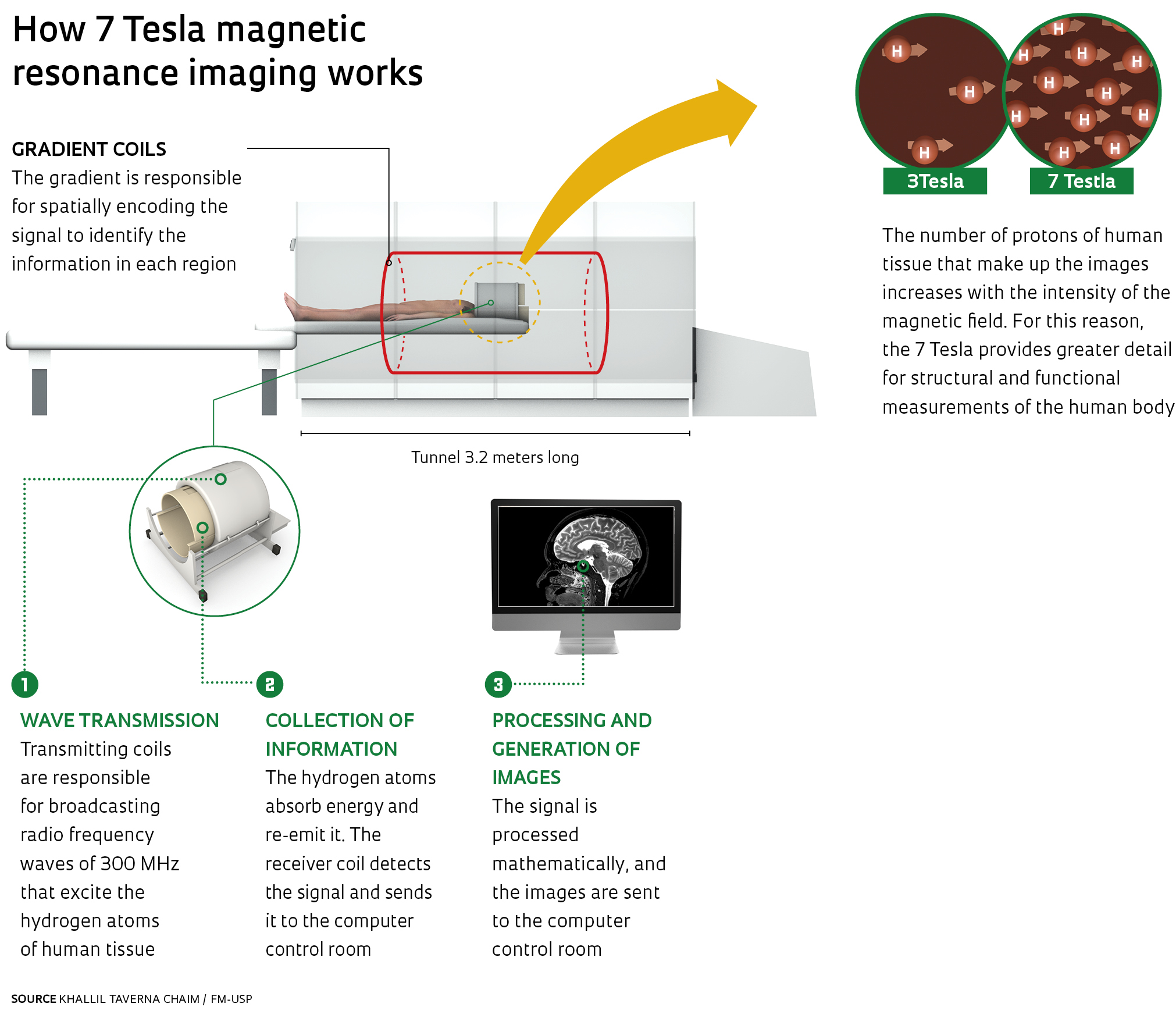
 The cost of the equipment was $7.695 billion and involved funding from FAPESP, USP and the Medical School Foundation. Made in Germany and England, the Magnetom 7T MRI is an ultra-high-field piece of equipment that provides a higher level of sensitivity and detail for structural and functional measurements of the human body using magnetic resonance imaging. Its diagnostic imaging technology helps to identify the properties of substances in the human body in a non-invasive manner. Using electromagnetic waves, the unit’s coils interact with tissues inside the body. Images are then constructed, by decoding the signals received from the hydrogen atoms of the water making up the human body. Tesla (named after Nikola Tesla, the inventor who made major contributions to the use of electricity and magnetism) is a unit of measurement of the magnetic field. The clarity of the images generated by a 7 Tesla unit, in terms of the resolution and the ability to discern changes, is more than 5.4 times greater than 3 Tesla equipment and 21 times greater than the 1.5 Tesla equipment used in hospitals. A two-fold increase in the magnetic field quadruples the sharpness of the images. The standard 7 Tesla has not yet been released for clinical purposes, but it is already being used in various research centers around the world. The Magnetom 7T MRI was acquired under the Multi-User Equipment Program (EMU) of FAPESP, aimed at buying state-of-the-art equipment as it becomes available for a large number of researchers, institutions in Brazil and even abroad, whose projects are selected according to very strict criteria.
Initially, more than 20 research projects will benefit from the new facility—some of them are ongoing and are using images taken by computerized tomography equipment (CT) of the SVOC. The arrangement will also include ultrasound and X-rays. The CT equipment was purchased with funding from the Dean of Research under a project of the Integrated Autopsy and Imaging Research Center (NUPAPI). One of the most ambitious projects is perhaps the Brazilian Imaging and Autopsy Study (BIAS), coordinated by Saldiva, which seeks to find alternatives to invasive autopsies using diagnostic imaging. The work of validating new methods will be based on comparative studies. With the consent of the family of the deceased, the strategy is to subject the cadaver to an MRI and then to a conventional autopsy, and then compare the results of the two procedures. One of the international projects to which the equipment will lend support is known as the verbal autopsy, a computer program that seeks to clarify the causes of death of an individual with a set of questions for the family. “It is a resource that is being used in remote places, where there are no doctors to verify the cause of a natural death,” Saldiva explains. The results of this questionnaire will also be compared to the magnetic resonance images and to the conventional autopsy, to measure to what extent they help determine the cause of death.
 Saldiva says that the Ministry of Health plans to expand the supply of death verification services in Brazil, with the goal of having one per 3 million inhabitants. “One limitation is a shortage of pathologists.” he says. “Performing autopsies is not a very attractive job for doctors: it requires extensive study, the work takes time and does not pay well.” Improving the quality of care through imaging techniques would help alleviate the problem. “There are more CT scanners than autopsy rooms in hospitals, and it is common to have more radiologists available than pathologists,” adds Saldiva. Researchers will not start from scratch. This work is being done using computerized tomography available at the SVOC, where 900 post-mortem examinations have been performed, 300 of them with angiography of the entire body, a technique in which a contrast fluid is injected into the bloodstream of the cadaver. This provides evidence to help determine the cause of death.
Comparative studies, Saldiva notes, can help to improve hospital quality. “A survey done on the accuracy of death certificates showed a 20% discrepancy rate, that is, the cause of death indicated in 20% of the cases was not the actual cause of death. The knowledge generated by the PISA platform can help determine if the hospital care was everything it could have been for the patient who died.”
 Ongoing research projects that will benefit from the new platform include studies of cardiovascular, pulmonary, oncological, neurological and obstetrical diseases, as well as research on advanced imaging techniques. “What all these projects have in common is that they involve post-mortem images and validation of microscopic and macroscopic diagnostic techniques,” says Edson Amaro Júnior, a professor with FMUSP’s Radiology Department and a member of the management committee of the initiative. The PISA project team will work in partnership with researchers from the United States, England, Germany, the Netherlands and Israel, which have formed a virtual interconnected global network. Collaborators include, for example, Kamil Uludag, a professor with the Cognitive Neuroscience Department of Maastricht University in the Netherlands, whose laboratory also works with brain imaging using the 7 Tesla MRI equipment. Or the German professors Waldemar Zylka, at Gelsenkirchen University of Applied Sciences, who has long collaborated with USP, and Harald H. Quick, at the University of Duisburg-Essen, one of the first centers to use full-body 7T equipment. Peter Morris of the University of Nottingham in the UK is one of the research partners of USP’s Physics Institute in São Carlos and of the University of Campinas (Unicamp) in the development of coils for 7 Tesla equipment.
Ten years ago FMUSP established what would become the world’s largest brain bank, with more than 3,000 organs. About 350 are collected every year through donations. In October 2014, the German neuroscientist Helmut Heinsen of the University of Würzburg, came to Brazil to work at the brain bank for two years. He uses a technique that dips the brain into a substance derived from cellulose called celloidin, which acquires a plasticized consistency. It is then sectioned into slices of less than 1 mm in thickness, which is used in studies of degenerative and neurological diseases. This project will also have an interface with the PISA platform: before sectioning, the brains will undergo 7 Tesla resonance imaging, and these images will be compared to those obtained after using celloidin and sectioning.
The project will include other aspects, such as medical education. “The impact of these images on the training of doctors will be great, at a time when the FMUSP curriculum is being revised and there is an emerging convergence between pathology and radiology,” says Amaro Júnior. The production of teaching materials, such as new atlases of anatomy, and the ability to compare images of healthy and diseased organs or tissues promise to improve the training of medical professionals.
The laboratory plan is designed to enable all the envisaged activities. Next to the reception area, there are two small rooms, intended for interviews with family members of the deceased to gather information and obtain consent for participation in research (see Infographic). Next to another entrance, there is a room for preparing the cadaver. Alongside the MRI equipment room there is a space for animal experimentation—panels installed in the wall, constructed so as not to compromise the room’s insulation, will exchange data with external experiments.
The facilities also have a larger space for training—which can be used for classes—a control room and various other rooms to house support equipment, such as air conditioners and chillers, devices that provide continuous ice water to cool the helium gas and other instruments used in conjunction with MRI equipment. Helium has to be kept in a liquid state at minus 269 degrees Celsius, to ensure the superconducting properties of the unit’s coils and generate the magnetic field.
The idea for the PISA platform began to emerge in 2009, when Saldiva and Amaro, in casual conversation, floated the idea of working together doing research using cadaver images. Saldiva took it upon himself to go to the FMUSP administration and ask for any available CT scanner that was being deactivated for use at the SVOC. And he got it. He then presented a plan to the Multi-User Equipment Program for acquisition of a modern MRI machine, one with a 3 Tesla field. FAPESP approved the project. The interest of several university groups in participating in the initiative led to a reassessment of its scope—and the idea of working with 7 Tesla equipment. “We asked for larger contributions from USP and the School and then things began to fall into place,” recalls Amaro Júnior. An agreement between FAPESP, FMUSP and the Medical School Foundation was signed in 2012.
In May 2012, the architectural design of the platform was determined, on land that served as a parking lot and pedestrian walkway behind FMUSP headquarters. Because it is a specially designated area, the decision was made to build an underground laboratory, which would take a year to be built according to the schedule provided by Siemens. “We held weekly meetings in order to keep the work on schedule,” says Marina Caldeira, FMUSP’s innovation manager and responsible for project monitoring. A management company was hired to monitor the construction and some changes to the design were necessary. The PISA platform facilities are next to the SVOC, and the idea was to connect the new laboratory to the underground tunnel linking the Hospital das Clínicas to the SVOC, where people who die in the hospital are taken after death. They discovered that the tunnel was much closer to the surface than anticipated, and the plan had to be revised.

Léo Ramos
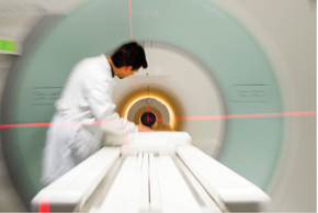
The Magnetom 7T MRI: manufactured in Germany and England by Siemens, has not yet been authorized for clinical useLéo RamosWhile construction of the building was underway, the FAPESP import division organized the procedures for purchasing the equipment, one of the most expensive purchases ever made by the Foundation. The task of purchasing the equipment and bringing it to São Paulo was coordinated by Rosely Aparecida Figueiredo Prado, affectionately known as Rose, the import and export manager of FAPESP. Contract negotiation, conducted during the second half of 2012, lasted a few months. “Some Siemens contract clauses did not apply to an institution like FAPESP and had to be modified,” says Prado. The formal start of the process occurred on November 12, 2012.
The equipment was manufactured by Siemens in two countries: the magnet came from England and the resonance unit from Germany. The challenge was to try to coordinate the timeframes for manufacturing and delivery of the equipment with the laboratory facility construction schedule. Pardo wanted to ship the two parts of the equipment on the same vessel, but this proved unfeasible.
The two ships carrying the equipment arrived at the port of Santos within a few days of each other. While the German unit set sail on October 6, 2014 and arrived at Santos on October 23, 2014, the magnet left England on October 2, 2014 and landed on October 29, 2014. On November 3, 2014, the cargo was cleared for import, but it was decided to store it for a few more days in the warehouse of the Deicmar company in Santos because the insulation of the room where the equipment would be assembled was not yet installed.

SIEMENS AND OTHER PHOTOS PROVIDED BY THE PISA PROJECT
The sequence of events: the equipment arrives at the port of Santos; its arrival at FMUSP; insulating the examination room with silicon steel; three tense moments hoisting the machine over the ceiling for installation in the laboratory; placement through the ceiling; and final finishing of the room SIEMENS AND OTHER PHOTOS PROVIDED BY THE PISA PROJECTA few months before the arrival of the equipment the process of importing raw material for the insulation began. It included special plates of copper, rock wool and silicon steel. Its supplier was the U.S. company ETS Lindgren, at a cost of $123,000. To expedite shipping, it was decided that all the material would be brought in by air. On November 8, 2014, four trucks went up the mountain with the disassembled resonance equipment and delivered it to FMUSP. A big test would come that day: the 2014 drought in Sao Paulo helped in the construction of the laboratory, but the first big rain put the drainage system to the test. The water moved toward the platform, but it was contained and the problem solved. Four days later the insulation material landed at Viracopos Airport and arrived at the university.
The hoisting of the Magnetom 7T MRI by crane took place on November 25, 2014. Since there is little space to maneuver around FMUSP, it took two cranes to lift the machine and place it within the platform through an opening in the ceiling, which was later closed. At every step of the process, those involved would discuss the difficulties that lay ahead, and Saldiva ended the conversation with a prayer: “Let us pray to the Virgin Mary, the Untier of Knots.” On hoisting day someone remembered to put a picture of the Virgin, an object of worship in a German church for over 300 years, inside the resonance equipment. On the eve of the inauguration of the platform, Saldiva commented that while the journey had been a long one, everything had worked out in favor of the initiative. “Everyone we showed the project to were supportive and agreed the idea was good. Instead of putting up obstacles, they proposed solutions. This almost never happens,” he says.
Project
Imaging Platform in the Autopsy Room (No. 2009/54323-0); Grant mechanism Multi-User Equipment Program; Principal investigator Paulo Hilário Saldiva (FMUSP); Investment R$10,352,243.31 (FAPESP).