NIBSC / SCIENCE PHOTO LIBRARY
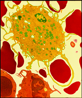
Capture: a defense cell called a macrophage (yellow) creates extensions and surrounds a red blood cell, before digesting it.NIBSC / SCIENCE PHOTO LIBRARYEvery cell in the body is like a bustling metropolis where, instead of cars and people, essential molecules and structures of varying sizes and types are continuously circulating to keep it alive. All this sometimes frenetic movement occurs in a limited space, defined by an extremely thin and pliable structure: the cell membrane. Formed of a double layer of lipids, a type of fat that gives it the viscosity of a light oil and makes it relatively fluid, this cell lining contains embedded proteins here and there. In recent years, our understanding of the membrane has increased. While fragile-looking under a microscope, it plays a much more complex function than just separating the contents of the cell from the external environment.
“The membrane is much more than packaging enclosing the cell’s content,” says biologist Bruno Pontes, a researcher at the Federal University of Rio de Janeiro (UFRJ). Currently at the Institute of Mechanobiology at the National University of Singapore, in Southeast Asia, as part of a postdoctoral internship, Pontes is part of a team in Rio de Janeiro investigating the physical characteristics of the cell membrane and recently measured its elastic properties with an unprecedented level of precision.
Coordinated by physicist Herch Moysés Nussenzveig, internationally known for his studies in optics, the UFRJ group includes biologists, mathematicians and, of course, physicists. In a series of tests carried out in the last few years, researchers used a very concentrated laser beam to manipulate the membranes of brain cells, blood cells and the cells of other tissue in the laboratory. Using these optical tweezers—at the brightest point, the laser induces electric dipoles, which can be used to attract and move microscopic objects and, for example, manipulate living cells without damaging them—they found that different types of cells have membranes with different elastic properties.
In very delicate experiments, Pontes used laser tweezers to trap microscopic spheres of a plastic material and then make them adhere to different points on the cell membrane. Seconds later, he pulled each of the spheres at a constant speed until the membrane stretched into an elongated tube—the force necessary to stretch the membrane and form a tube is on the order of tens of piconewtons, a few trillionths of the force that gravity exerts on an apple.
Measuring the radius of the tube and the force needed to form it, he was able to calculate the two physical quantities that determine the elasticity of a membrane: the surface tension (resistance to breaking) and bending stiffness (resistance to bending). In some cases, the elasticity of the membrane varied so much from one cell type to another that, according to Nussenzveig, “it seemed obvious that there must be a direct relationship between these membrane properties and the function the cell plays in the body.”
Neurons, the most abundant cells in the brain, responsible for the storage and transmission of information, were also the cells with the most flexible membrane of the five cell types evaluated. With a specific, characteristic shape, the neuron has a bulkier region, the cell body, which contains the nucleus, and another composed of narrower, elongated extensions, called axons and dendrites, through which electrical signals travel to reach the next neuron. In the brain, one neuron connects to others through these extensions, which can be constantly reshaped. Since they maintain this plasticity and are quite asymmetric, it makes sense, according to the UFRJ group, that their membranes are more malleable.
The second most malleable were astrocytes, according to the results that the researchers published in July 2013 in the journal PLoS One. Astrocytes look like stars and are the second most abundant cell type in the brain, where they play essential roles in nourishing neurons and regulating the formation of synapses, connections between one neuron and another.
Interestingly, the brain cell with the most rigid membrane is also usually more active and is able to undergo greater deformation: microglia. Similar to astrocytes, but with longer extensions, microglia are the main central nervous system defense cells. With these extensions, they constantly probe the environment looking for diseased cells and infectious agents. When they find them, they create extensions and encompass them in order to then destroy them in a process called phagocytosis.
In the researchers’ interpretation, it makes sense that the physical properties of the membrane vary according to the cell type. After all, different cells perform different functions in the body. “The membrane is the interface between the cell interior and the external environment, allowing interaction between them,” recalls the physicist, coordinator of the UFRJ Optical Tweezers Laboratory. “It also detects chemical signals and mechanical stimuli in the surrounding environment and transmits them to the inside of the cell. At the same time, it serves as a platform for a cell to transmit signals to the rest of the body, indicating, for example, the need to produce antibodies. Furthermore, the membrane gives the cell shape, and also deforms, allowing the cell to move by creating projections,” he concludes.
In experiments done at UFRJ, Pontes and other researchers on Nussenzveig’s team also found that the membranes of microglia have the same elastic properties as the membranes of another defense cell: macrophages, which are produced in bone marrow and released into the bloodstream, through which they spread throughout the body (except for the central nervous system). In a manner similar to microglia, macrophages also perform phagocytosis, emitting extensions that identify, encompass and destroy old cells, infectious agents and particles foreign to the body.
 According to the UFRJ group, a common embryonic origin could explain the fact that the membrane of macrophages and microglia share the same elastic properties. Both cells are derived from the mesoderm, one of the three layers of cells that form the embryo in its early stages (other cells of the central nervous system originate from the ectoderm). They retain many common features, despite migrating to different regions of the body during development—microglia travel to the central nervous system, while macrophages circulate through peripheral tissues.
“They are like brothers who were raised together in childhood, but as adults went to live in different countries,” describes Pontes. “They preserve many common features, though they are located and function in different contexts.” Nussenzveig reminds us that both microglia and macrophages have to withstand strong forces and large surface deformation during phagocytosis, which would justify more resistant membranes.
This stiffness, however, is not permanent. It is about four times greater than that of the membrane of neurons when the microglia and macrophages are inactive, in a dormant state, and falls to about half of the initial value when these defense cells are activated.
The researchers recorded this increased flexibility when they treated the macrophages and microglia with compounds found in the walls of bacteria. These compounds arouse defense cells, making them active. “The decrease in bending stiffness allows these cells to bend and issue extensions, preparing to engulf other cells” explains Nussenzveig.
The similarity found between macrophages and microglia was also observed between astrocytes and glioblastoma cells, the latter being a devastating type of brain tumor that results from the uncontrolled proliferation of astrocytes. “We do not know the details of how these properties influence the function of a cell,” says Pontes. “But the fact that the elastic constants change according to the environment and the state of the cell certainly exerts some influence on their performance,” says the biologist, who works in Singapore with Nils Gauthier’s team, trying to better understand how these elastic membrane properties might orchestrate a series of phenomena within the cell.
“This is fairly consistent evidence that the elastic properties of the membrane have a direct relationship with the cell’s function in the body,” says Nussenzveig. In the article published in PLoS One, the team at UFRJ also demonstrated that the flexibility of the membrane does not depend only on the lipids in it. What largely determines its stiffness is called the actin cytoskeleton: a diffuse network of filaments of actin protein distributed throughout the cell and anchored in the proteins trapped in the membrane.
Prior to this study, it was believed that the membrane tubes that are formed when the cell is manipulated with optical tweezers were made of pure membrane, or almost exclusively lipids. The group at UFRJ demonstrated that, when pulling the membrane the cytoskeleton is also dragged along, together with the lipids. Previous observations, performed by the group of Michael Sheetz, director of the Institute of Mechanobiology in Singapore, where Pontes has been undertaking his postdoctoral research since the beginning of 2013, did not take into account the influence of this protein fiber network. This situation, according to the researchers from UFRJ, does not match reality. “A cell with a pure membrane, uncoupled from the cytoskeleton, can’t exist because it would be very unstable,” explains Nussenzveig. “In the cell, the membrane is anchored in a kind of actin mat, the cortex, which gives it greater stiffness.”
His group also found that the cell membrane is ten times tougher than imagined. An optics specialist and creator of the UFRJ Optical Tweezers Laboratory, he and the physicists Nathan Bessa Viana and Paulo Américo Maia Neto realized that, in general, the optical tweezers—which consist of a laser system coupled with a microscope—suffered from a kind of defective vision, which interferes with measurements. This defect is an optical aberration called astigmatism; a change in the focus of the laser decreases the force it can exert. After studying the issue for 13 years, the team at UFRJ claims to have finally identified the cause of the problem and found a way to fix it. “The paper describing these corrections has been submitted and should be published soon,” Nussenzveig says.
“Finally, the tweezers have been fully understood from first principles,” says the physicist. He believes that his group has now managed complete control over the tweezers and how to increase their capture power. “Until our work, calibration was done indirectly by comparing with hydrodynamic forces, caused by the friction between a plastic microsphere and a fluid,” he says. Major differences were seen, on the order of a factor of 10, in the measurements made by different laboratories as a result of the less precise calibration of the instruments. “Our group is the only one so far to obtain absolute calibration and our results are reliable within the precision that is achievable in cell biology,” says Nussenzveig who, at 80, remains enthusiastic about his research and knows that we are far from achieving a physical model of the cell membrane. “There are theories that seek analogies with materials that we understand to describe the functioning of the cell membrane,” he says. “But they are rudimentary. It is not enough to treat the materials like inert, passive systems. You must take into account the reactions of the cell as a living system.”
Scientific article
PONTES, B. et al. Membrane elastic properties and cell function. PLoS One. July 3, 2013.