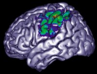
brain institute/iiepPlaying with words… Seeing if bread and red rhyme activates concentrated areas of thebrain institute/iiep
The moral and human facility of making moral judgments or emitting important opinions have historically been considered themes appropriate to philosophy, to the point of having constituted from the time of Nicomachean Ethics, by Aristotle, an almost autonomous field of philosophy. It was more or less this that a young neuroscientist heard from the reviews of a respected international publication when he attempted to register his first article, with results obtained via functional magnetic resonance scans, suggesting that the front polar cortex and the anterior temporal of the brain were activated when healthy volunteers set about a determined task that involved moral judgment. It was recommended to him that it would be better to leave neuroscience out of these complicated meanderings of ethics for which only philosophers had always demonstrated the most explicit aptitude.
At that time, approaching 30 years of age and confident in what he was doing, the young researcher clearly did not follow the suggestion. Instead of that, he reorganized the article, whose authorship he shared with both a Brazilian and an American colleague, and sent it to the respected magazine Arquivos de Neuropsiquiatria [Neuropsychiatry Archives], edited in Brazil, but with international circulation guaranteed by way of the SciELO, the Scientific Electronic Library On-line. And it was in this manner that the conclusions of the study carried out by Jorge Moll – and this is the name of our personality – , presented in a pioneering manner in 2000 to the American Society of Neurology, had finally appeared in the Archivos [Archives] in an article signed for by him, as well as fellow authors Paul J. Eslinger and Ricardo de Oliveira-Souza, with the title, “Frontopolar and anterior temporal cortex activation in a moral judgment task”, in July of 2001. Coincidentally, the publication occurred in the same month in which there came out in the international magazine, which he had originally sought out, an article from a Princeton University group led by the philosopher Joshua Greene, concerning the direct involvement of emotions in decisions that implicate major moral dilemmas.
The strategy of Moll of not putting off the publication of the article linked to a theme that he knew had a lot of appeal, and sending it shortly afterwards to a theoretically lesser known magazine, showed itself to have been correct in order to formally place it, without further delays, onto the international scenario in his field of research: during 2000 he published in Neurology his first study in which, during the period from 1997 to 1999, he made more use of the deep images of the brain obtained by way of functional magnetic resonance, a technique that, in simple and synthetic terms, captures images of the brain during activity. Various others have followed in different publications, such as NeuroImage and The Journal of Neuroscience, until the important revision article on the neurological fundamentals of moral judgment entitled, “The neural basis of human moral cognition”, signed for by him and Roland Zahn, Ricardo de Oliveira-Souza, Frank Krueger and Jordan Grafman, published in Nature Reviews Neuroscience in October of last year.
Moral brain
During these few years of consistent affirmation of the researcher’s work, always starting from functional neural images, Moll has arrived at various conclusions with huge potential for producing polemic situations. For example, that in which the frontal lobe of the brain, home of the executive functions according to the most respected current theories, and which maintains itself firmly engaged in the solution of difficult and complex problems by way of rational logic, is also involved in the most simple and routine tasks. “This that in this lobe can be identified as our moral brain is in truth an ongoing sensor active, untiring, involved with the most simple things, such as, for example, deciding whether or not to sleep a little more, and not only with the judgment of major issues, such as the launching of an atomic bomb over Hiroshima”, he says. Indeed, the capacity to store a large repertoire of situations and to perceive them in their multiple connections would allow this moral brain, among other things, to anticipate the pathways to take much before acting.
Another of the researcher’s conclusions, whose correlated scientific article is in the publication phase, indicates that an absolutely identical area of the frontal-mesolimbic system is activated when a person aiming only financial rewards or looking at moral reward even at the cost of financial damage. In the study that produced these results, done with the support of very high electromagnetic field functional images and during which healthy people had to make real decisions to earn or lose real money (up to US$ 128), the volunteers received a list of philanthropic organizations of all ideological matrices, among which were some in which both the organization chosen and the investor would gain something, others in which only the investor would gain and others in which, in the case of investing money there, he would lose the corresponding amount.
The example of Dr. Moll, today 35 years of age and a researcher linked to the Cognitive and Behavior Neuroscience Unit of the D’Or Network, a private institution located in Rio de Janeiro – but at the moment based in the United States, where he is carrying out his post-doctoral studies at the National Institutes of Neurological Disorders and Stroke, linked to the National Institutes of Health (NIH) –, is highly illustrative of what has been occurring in the country, in the environment of neuroscience studies based on the technologies of neuro-image, especially those produced by functional magnetic resonance. With the help of these images that provide some very precise temporal indications of the brain at work, researchers have sought to discover, among other findings, pari passu with his colleagues from the more developed countries, the physiological correspondence in the neurons of language acts, movements; moral judgments; emotions; sentiments, social behavior etc., put into effect by healthy people. And has certainly contributed to making an advance in the current knowledge about the brain, at the same time that it indirectly proposes new means for the clinical coverage of important neurological illnesses.

brain institute/iiepTo create words starting from whatever letter brings more dispersed areas into actionbrain institute/iiep
Nevertheless, in a field so vast and swampy as much as is the expressiveness of the particular human being in each man and woman, there are criticisms, obviously, to what would be the strict limits and the character, very much relative, of the information and conclusions that are seen to elucidate it based on the functional neuro-images. And these criticisms, begin within the neuroscientists’ own midst, only to later extend to those who study the humanities of varied shades. “This elegant manner, because it’s not invasive, of attempting to catch the functioning of the brain has some problems, and it all begins because it happens like a mode of castle blocks”, says the neurologist Fernando Cendes, head of the Neuro-imaging Laboratory of the Medical Science Faculty of the State University of Campinas (Unicamp).
“There is always an ‘on’ condition and an ‘off’ condition”, continues Cendes, “that are investigated with the individual inside the magnetic resonance machine, and after subtracting one situation from another in order to verify what was more active in the brain in the ‘on’ situation” Or that is to say, the task of moving the fingers of the hands would be an ‘on’ situation, and next, remaining still would be an ‘off’ situation. “Now, the finer and more complex that one wants to investigate, for example, the cognitive process or the thinking processes, then it’s much more difficult to propose the ‘off’ condition. It’s not possible to seriously propose to a volunteer, for example, not to think”, he comments. Another problem is that normally the volunteer within the magnetic resonance tube cannot speak (he usually responds yes or no by managing a joystick) and can almost not move beyond the limited movements that he is instructed to do.
Redundant activity
Cendes, a specialist in epilepsy, also observes that there is lots of redundant brain activity, hence or a determined process, such as the cognitive, activates areas in a very similar manner to another, such as that of attention. And it is precisely by reason of this redundancy that, in determined pathological circumstances, large areas of the frontal lobe can be removed without lots of damage, because another area takes over the functions linked to those that were lost. “To verify which area is activated during the execution of a task is OK, but one can’t guarantee that it’s only and exclusively that area which is activated during this moment”, he observes. For this reason one must treat all of the studies based on functional magnetic resonance with a certain amount of caution, because, he says, when faced with the brain “We’re still facing a jigsaw puzzle full of challenges, in which nothing is as simple as it may seem.” Anyhow, the research based on functional images definitely has advantages, he adds. “They’re interesting, important and have been adding knowledge on the working of the brain.”
Cerebral atlas
A contribution to this end has been offered by a research group originating from different institutions and now brought together at the Albert Einstein Israeli Teaching and Research Institute (IIEP), under the direction of Carlos Alberto Moreira Filho. It is worth recalling at this point that the IIEP, linked to the Albert Einstein Israeli Hospital, is a private partnership of two state universities and one federal university located in the state – the University of São Paulo (USP), Unicamp and the Federal University of Sao Paulo (UNIFESP) – within the CinAPCe program, which alluded to cerebral synapses and is an abbreviation in Portuguese for Inter-Institutional Cooperation to Support Brain Research. This initiative, which has FAPESP’s financial support and should effectively enter into operation by the beginning of 2007 (see Pesquisa FAPESP, issue 124), could provide an impulse towards advances in this research area that over the last few years has clearly been building up a certain force in the country.
At this point in time, the IIEP, by way of its participation in the CinAPCe, has already acquired, with its own resources, one of four magnetic resonance machines of high electromagnetic field with which this brain research network, whose first focus of investigation will be epilepsy, will be able to count upon. They are machines with an electromagnetic field of 3 Teslas, which allow for obtaining cerebral images with a definition and spatial resolution much greater than those offered by the machines in use today, those of up to 1.5 Tesla, and in lesser time than the patients spend in the current machines.
But, even before the new machine was installed, the IIEP has been working with the available equipment in a series of research projects that make use of both functional resonance and a combination with other techniques for obtaining cerebral images. And, in some cases, there is even a strict articulation of research interest with immediate clinical objectives. An example is obtaining images that allow for reducing the risk of hitting areas linked to important functions during surgery for the resection of brain tumors. “Shortly the teams from the IIEP and the Albert Einstein Hospital will initiate a protocol in which the functional resonance image will be integrated with a navigator in the surgical field, thus allowing the realization of very high precision surgeries”, says Moreira.

brain institute/iiepPlaying with movements: moving the fingers of the left and right hands mobilizes neurons of different areas of the cerebral cortexbrain institute/iiep
The dream is free and unlimited, but, in order to stay on more concrete land, one of the IIEP relevant projects based on functional magnetic resonance is the mounting of a data bank aimed at the creation of a type of cerebral atlas of the population who seek out the hospital, a functional normative base that will permit an unlimited set of comparative studies of healthy cerebral conditions with pathological conditions. In order to begin this data bank, which should be shared with those of other countries, until this moment 50 healthy volunteers, men and women between the ages of 18 and 54 years, previously submitted to a battery of psychological questionnaires, have made themselves available to remain almost one hour inside the magnetic resonance equipment carrying out simple tasks capable of activating areas of the brain linked to motor, visual activities, verbal fluency, tactile sensibility and specific language (rhymes, semantics, among others).
Given that each task carried out inside the apparatus generates almost 3,000 images, as the coordinator of this image bank explained, the radiologist Edson Amaro Júnior, a professor at USP’s Medical Faculty and a researcher with the IIEP, and the biomedical doctor Maria Angela Barreiros, who works with him, and with the 50 volunteers who up until now represent the bank, they already have a total of 750,000 images obtained by functional magnetic resonance, which will allow for producing 720 maps of cerebral working.
This means abundant material to be worked upon in the so called post-image, the phase in which there is refinement of the highly smudged information using computer tools, say, captured via the magnetic resonance equipment and based on the differences of oxygenation of the cerebral areas under examination before and during a task – that story of the ‘off’ and ‘on’. Within this environment there is an important study being developed at the IIEP under the coordination of the computer scientist Griselda Jara Garrido, who in different projects is working on the development of methodologies that allow for a more precise interpretation of the cerebral images, for the articulations between data banks and other sources of information.
One of these projects is the setting up of a neuro-informatics laboratory with a strategy of administering knowledge based on the web and high performance computing. “We’re making use of the availability of free tools to mount a laboratory that, by the end of 2007, will have a cluster of 8 nodes for the parallel processing of images”, explains Griselda. In simple terms, she explained that all the effort of the part in which she is working is destined to making explicit what the image generated by the resonance equipment gives out so that this data can be interpreted by neurologists. We can say that we are dealing with a fundamental half way field between radiology and neurology.
The young researcher is participating with colleagues from the University of Western Australia in a study that is demonstrating that there are sensitive structural effects in the brain coming from the continued use of cigarettes, a study sent for publication in Neurobiology of Aging. With the support of a young researcher grant conceded by FAPESP, she is also one of the authors of a study into anti-social behavior being developed at the Cognitive and behavior Neuroscience Unit (UNCC) of the D’Or Labs-Hospitals, being coordinated by Jorge Moll. Griselda observes that the so called anti-social behavior only merit medical and psychological attention when it becomes recurring, chronic, “and causes problems in multiple spheres of life”. And it is then that the expression “anti-social behavior disorder” is applied or ASBD.
Knowledge based on illness
Research with neuro-images became fashionable at the beginning of the decade of the 1990s. But when including among the fundamental techniques for the recent advance of knowledge about the brain Positron Emission Tomography (PET), one can back date the starting period to the previous decade.
Nevertheless, it is worth while going back much further until one arrives at the twenties of the previous century, in order to perceive how in truth the studies on the healthy brain, with the support of techniques such as functional magnetic resonance, have their roots in the research of lesions, and especially in knowledge generated during surgeries that attempted to calm down forms, more or less serious, of epilepsy.
“When a patient went to the epilepsy operating theater he had his brain stimulated by a low ampere electric current because it was necessary to verify which areas had been linked to speech, to movement etc. This was a situation in which this stimulus was absolutely necessary, since the surgeon was going to remove a large area of the brain and was looking to preserve to most important functions”, says Fernando Cendes, the head of the Neurology Laboratory at Unicamp and one of the most respected specialists on epilepsy. Decades of experience with these complicated circumstances ended up producing a cerebral mapping that was extremely trustworthy.
And even today, during surgery, or in the placement of an electronic plate, electrical stimulus is the safest means for this necessary preservation of functions. Another pathway, later on, which led to a greater understanding of the brain, was the observation of the damages of a heart attack or a vascular cerebral accident (VCA, the well known stroke) and the correlations established between the functional deficits that the patient presented and that which was perceived in images, observes Cendes.
The researcher has no doubt, indeed, that it was the investigation of epilepsy, a condition that can be caused by an enormous number of different factors, from VCA to genetic causes, passing through tumors, the majority responsible for the advance of cerebral research in Brazil. “And it’s not just in the country. This is traditional throughout the world: the centers that lead with epilepsy always have highly qualified people also working with cognitive functions, because by way of clinical necessity they’re going to be colliding with, in the best sense, the advance of research.” Fernando Cendes observed that Brazil is an advanced country, of the state of the art, as is said in the media, in relation to epilepsy surgery groups.
“Without false modesty, we’ve good insertion in international terms and we owe nothing to the more developed countries”, he says. The technical scientific conditions of the various brain research groups are also very good. But, as in almost all of the country, the counterbalance to this is the enormous number of unprepared doctors in the health system and dramatic problems such as the incidence of highly avoidable illnesses like neurocysticercosis. “We have limitations that border on the ethical, in the manner of what to do to use equipment for studies, if it’s the same equipment for the diagnosis of patients – In this case the researcher works at the weekend and at night.” Even at that, the diagnosis is: the area of neuroscience is strong in the country and is tending to advance considerably with programs such as the CInAPCe.
The study in which she participated followed up 15 people with ASBD, with a control group of healthy people within the same age range, gender and schooling, and made use of a method that allows one to obtain measurements of the density and volume of the grey matter (voxel-based morphometry). Clearly the analyses for the region of interest, based on functional magnetic resonance, was also used. And what was found, according to researcher Griselda, was that “the results confirm the hypothesis that a network of cerebral regions involved in the mechanics of moral cognizance and moral emotions is intimately related to the genesis of anti-social behavior in the psychopathic personality”. 3-dimensional images of the brains showed that there was coincident activation of these areas.
The Brain Institute of IIEP is in truth, in spite of its still short time of life (it was established in 2003), the home of various other research projects, involving varied combinations of techniques of obtaining cerebral images. If there is a concern in creating a reference normative data base of healthy people, on the other hand cancers, Parkinson’s disease, Obsessive Compulsive Disorder (OCD) are some of the illnesses focused by the Institute’s researchers. And if functional magnetic resonance is an important tool, structural magnetic resonance, the electroencephalogram, a combination between these techniques, and new ideas such as diffusion tensor imaging, have also considerable weight in the efforts of the IIEP to be included among the centers of advanced brain research reference in the country over the next few years.
Even though São Paulo is the most advanced center for brain studies in the country and has the best infrastructure for this type of research –“there’s more magnetic resonance on Paulista Avenue and its surroundings than in all of the Latin America Countries put together”, comments Fernando Cendes. Research centers involved in this field distribute themselves through Porto Alegre, Curitiba and Rio de Janeiro, although the Federal University of Rio de Janeiro (UFRJ) does not have a single magnetic resonance machine appropriate for research. Even with such limitations it is in this university’s laboratories, more precisely at the Neurobiology Department of the Biophysics Institute, where she is an assistant professor, Claudia Vargas works with brain activity maps, making use of functional magnetic resonance, of electroencephalograms and trans-cranium magnetic stimulation. She is very interested in the small variations of excitability of the neurons of the cerebral cortex, having managed to apply a brief magnetic field to the scalp of volunteers. And what does she want to verify with this?
With the application of these magnetic pulses to the so called primary motor cortex, the region of the brain that acts in the control of body movement, Claudia is managing to map out alterations in the level of brain activity associated to the movement of determined muscles and to identify, in these maps, changes brought about by problems such as the amputation of limbs. “There’s a representation of the movement of limbs in the motor cortex. And if a person loses a hand, for example, the representation of the arm expands to occupy that space that was for the hand”, she explains.
Neuronal plasticity
What appears extraordinary is that, when a hand transplant occurs, the muscles of the donor progressively gain representation in the motor cortex. Claudia was able to verify this when working, since 2002, with a research group at Lyon, France, coordinated by Dr. Angela Sirigu, who accompanies the two hand transplants carried out at the Edouard Harriot Hospital under the command of the surgeon Jean Michel Dubernard. The results obtained via functional magnetic resonance by this research group on the first transplanted patient were published in 2001 in Nature Neuroscience. “We made various tests with the patients, and what we think is that the reorganizations observed in the cerebral cortex after the transplant are possibly derived from the change of electrical activity of the neurons. This change, for its part, occurs thanks to the reconnection of the peripheral nerves and to the use of the new limb, results that reinforce the idea that plasticity exists in the adult brain”, comments Claudia, after telling that the group is currently completing the article concerning this part of the experiment.
In the same UFRJ department, assistant professor Eliane Volchan has been working on a research line baptized as ‘emotion regulation’, in which one attempts to verify when and how we manage to control our emotions. Yet again, it is functional magnetic resonance that is the basic tool for the studies, and therefore care is taken to verify the activation of determined areas on the cortex. In the most recent experiments that she carried out in collaboration with Drs. Jorge Moll and Letícia de Oliveira, from the Federal Fluminense University, and Luiz Pessoa, currently at Brown University in the United States, volunteers within the magnetic resonance tube carried out the simplest task of answering if two small white stripes upon a black background on a computer screen had the same direction or not, as quickly as possible. Simultaneously, sometimes neutral photographs, sometimes photographs with strong emotional content were rapidly shown whilst the volunteers performed the task of the little stripes, and the instruction was to pay attention to this task, without giving any importance to the photos.
When the aversive photo appeared, the volunteers took longer to identify the direction of the stripes, an indication that emotion interfered in the performance of the task. In a control situation, it was explained that the photos of mutilation were the result of make up work for the cinema. “It was verified that the interference of emotion in carrying out the task disappeared when the people were informed that the aversive photos were fictitious”, says Eliane. In her research Eliane has also been working with other parameters, with people being experimented upon outside of the magnetic resonance machine. Indications of blood pressure, heart beat etc. are used as a complementary form to images.
The studies with functional magnetic resonance have always been done, up until now, with healthy individuals, although it seems that they can say something about determined illnesses as well. And it is important to emphasize, at the same time, that the first basis for the studies with healthy individuals was provided by research of illnesses, especially that of epilepsy (see in-set on page 43). “When we worked with the difference between factual and moral judgments, we saw that two regions of the temporal lobe, one the anterior and the other the posterior, the so-called STS, famous in studies with monkeys and related in humans to complex perceptual stimuli, had been activated. Now this region is involved in a hypoactive manner in illnesses such as autism, in which the person deals with the others as object”, ponders Moll. Alright, possible interactions between health and illness, moral sense and the emotional part, what appears to be improbable is that some day the philosophers will believe that the notion of right or wrong is philosophically inscribed in the human brain.
Republish