
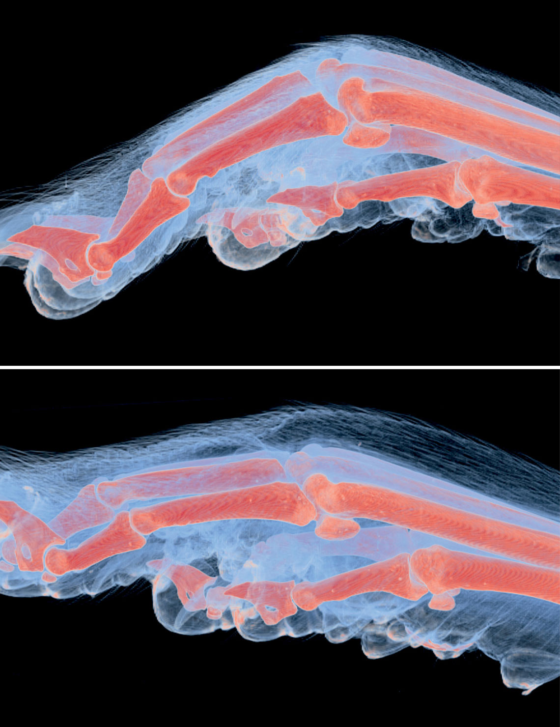
Ana Carolina de Carvalho / CNPEMThe paw of a healthy mouse and with swelling caused by the Mayaro virus (below), seen in microtomography images generated by the UVXAna Carolina de Carvalho / CNPEM
With the aid of three-dimensional images produced by the UVX particle accelerator, located in Campinas, in the interior of São Paulo State, Brazilian researchers are observing in detail how the Mayaro virus establishes itself in the body and the damage that it causes in different organs and tissues. Isolated for the first time in 1954 on the island of Trinidad and Tobago, in the Caribbean, the virus is transmitted to primates—particularly monkeys—via bites from mosquitoes of the Haemagogus genus, common in humid forest areas and transmitters of the yellow fever virus. Mayaro virus has already been detected in at least 14 countries of Central and South America, including Brazil, and causes a mild febrile illness that lasts for about a week in humans. Marked by headaches, muscle and joint pain, as well as the appearance of red rashes on the skin, Mayaro virus infection can, however, evolve to prolonged and painful inflammation of the joints, as debilitating as that caused by a related virus, the one that causes chikungunya fever.
In one of the laboratories of the Brazilian Center for Research in Energy and Materials (CNPEM), where the UVX operates, the group led by biologist Rafael Elias Marques inoculated small amounts of the virus into one of the back paws of mice and observed what happened over the course of a few days. The rodents had been genetically modified to be less resistant to viral infection and, therefore, were able to simulate infection in people who become more severely ill. The results, published in March in the International Journal of Molecular Sciences, revealed two types of effect: one local and another systemic.
In the first days, the virus caused intense swelling on the paw, which affected the skin and the muscles, ligaments, and joints. “The volume increased gradually, but suffered a more marked increase from the third day, when the paw was 50% larger than those of noninfected animals,” says biologist Ana Carolina de Carvalho, who is doing a PhD under the guidance of Elias Marques. The researchers attribute the swelling to inflammation caused by the replication of the Mayaro virus, which attracts defense cells to the location in which the virus reproduces.
Calculating how the volume of the swelling advances, according to Carvalho, was only possible with the use of potent X-rays emitted by the UVX, the first synchrotron light source in the Southern Hemisphere. The light generated by this equipment enables the visualization of structures of a few micrometers (thousandths of a millimeter) in size, at a resolution similar to the most powerful optical microscopes, but with important differences. This form of radiation passes through the skin and allows three-dimensional images of soft tissue to be created, without destroying the sample. “We managed to create a film of these structures in 3D and in high resolution,” reports the researcher. The UVX was deactivated in 2018 and replaced with Sirius, one of the most modern synchrotron light sources in activity (see Pesquisa FAPESP issue nº 269).
As it multiplied in the tissues of the paw, the Mayaro virus also spread rapidly through the body. Analyses of cells and tissues showed that, on the same day of the infection, there were already elevated amounts of the virus in the spleen and liver. The infectious agent took at least one day to reach the brain of the animals, according to the study, in which researchers from the University of Campinas (UNICAMP), the School of Medicine of São José do Rio Preto (FAMERP), and institutions in Germany and Belgium participated. On the fourth day of the experiment, all the infected animals had died.
“The work was very well done,” says virologist Pedro Vasconcelos, a researcher from Pará State University (UEPA) and the Evandro Chagas Institute, the center in which the Mayaro virus was identified in Brazil, in 1954. “The images show the effect of the virus in rich detail, indicating where it stays and what it causes in the joints,” says the researcher, who did not participate in the current study. However, Vasconcelos stresses that the infection spread more quickly in the animals than seen in human beings, since the rodents were genetically modified to have a less efficient defense system.
The team from CNPEM now plans to use the beams of light from Sirius to observe how the virus infects the cells. “In the near future, we want to use biochemical techniques to mark the molecules involved in the infection and, with the new synchrotron light source, observe the virus entering the cells and spreading through the body in real time,” says Carvalho. Using a microscopy technique capable of identifying biomolecular structures with a near atomic scale resolution (electron cryomicroscopy), Elias Marques’s group had already elucidated the structure of the Mayaro virus in 2021 and published the result in Nature Communications.
Since it was first identified in Brazil, in Belém, the virus has caused at least five outbreaks in human beings in countries covered by the Amazon forest, with around 1,400 suspected and a little over a hundred confirmed cases, with no deaths reported. It has also been detected in the state of São Paulo and, in March of this year, in Paraná, carried by people that were infected in areas of risk. Specialists in diseases transmitted by insects (arbovirus) fear that the Mayaro virus could adapt to the Aedes aegypti mosquito, which transmits the dengue virus, and is found in urban centers. According to Vasconcelos, laboratory tests have already shown that the virus is capable of infecting A. aegypti, something that has still not been seen occurring spontaneously in natural environments. There is no approved vaccine against the Mayaro virus, nor treatment for the infection caused by it, which tends to be confused with chikungunya.
Projects
1. Disease and resistance mechanisms involved in mayaro fever in mice (nº 18/03917-6); Grant Mechanism Regular Research Grant; Principal Investigator Rafael Elias Marques Pereira da Silva (CNPEM); Investment R$205,636.10.
2. Characterization and therapeutic potential of chemokines in flavivirus-induced sepsis and encephalitis (nº 18/10990-1); Grant Mechanism Regular Research Grant; Principal Investigator Rafael Elias Marques Pereira da Silva (CNPEM); Investment R$270,044.24.
3. Development of a first experimental Ilheus virus (ILHV) infection model in mice and design of therapeutic strategies (nº 18/02993-0); Grant Mechanism Doctoral Fellowship; Supervisor Rafael Elias Marques Pereira da Silva (CNPEM); Beneficiary Ana Carolina de Carvalho; Investment R$323,160.78.
4. Development of a usutu virus infection model in mice: Study of the disease and testing of a neuroprotective compound (nº 18/02594-9); Grant Mechanism Doctoral Fellowship; Supervisor Rafael Elias Marques Pereira da Silva (CNPEM); Beneficiary Rebeca de Paiva Froes Rocha; Investment R$274,749.47.
Scientific articles
DE CARVALHO, A. C. et al.Characterization of systemic disease development and paw inflammation in a susceptible mouse model of mayaro virus infection and validation using X-ray synchrotron microtomography. International Journal of Molecular Sciences. mar. 2, 2023.
RIBEIRO-FILHO, H. V. et al. Cryo-EM structure of the mature and infective mayaro virus at 4.4 Å resolution reveals features of arthritogenic alphaviruses. Nature Communications. may 24, 2021.