
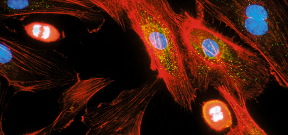
J. ZBAEREN/EURELIOS / SCIENCE PHOTO LIBRARYEndothelial cells: Record of the inflammationJ. ZBAEREN/EURELIOS / SCIENCE PHOTO LIBRARY
Cells have a memory. Possibly not all cells have a memory, but some are able to remember, later on, the conditions of the organism and the environment from which they were extracted. This ability to retain and transmit information to the descendants was not observed – as one might expect – in neurons, the brain cells that transport information in the form of electric signals from one point to the other of the organism and store this information in the brain. The team led by pharmacologist Regina Pekelmann Markus identified the cell memory in the endothelium, the layer of cells that line the interior surface of blood vessels.
So far observed in rats, this form of remembering, described in an article published in November in Plos ONE journal, is expected to arouse medical interest because of the power to influence organ transplants and the lab development of tissues that substitute natural tissues. “If the findings are confirmed in human beings, it will become necessary to pay attention to cell memory in order to obtain more homogeneous tissue cultures and reduce the risk of rejection in the case of transplants,” says the researcher from the University of São Paulo (USP).
The discovery of cell memory occurred quite unexpectedly. At the Chronopharmacology Laboratory of USP?s Biosciences Institute (IB), the group led by Regina was cultivating endothelial cells of healthy rats in acrylic recipients. The endothelial cells of animals submitted to a test simulating an acute infection triggered by the injection of LPS molecules from the bacteria’s cell walls were also cultivated in the same manner. After reproducing in vitro for nearly three weeks, the cells that descended from the cells removed from the rats behaved just like their great-great grandparents.
The cells extracted from rodents with the inflammation reproduced the physiological processes that occur in the endothelium of an injured part of the body: they attracted and retained the defense cells – especially the neutrophiles, the most abundant cells in the organism and one of the first cells to reach the inflamed region. The endothelial cells that stemmed from the cells removed from the rats without inflammation acted as if they were in a healthy environment.
If this phenomenon occurs in rats, the experimental model of various diseases, it is possible that it occurs in human beings as well, as the physiology and structure of human organs and tissues and those of murines are very similar. If cell memory is identified in human beings, this memory might explain, at least in part, the rejection of transplanted organs. Right after a heart attack, for example, the endothelium cells produce and expose molecules that attract neutrophiles to their surface. Normally dragged at high speed by the blood stream, the neutrophiles adhere to the endothelial cells that slow them down them until they stop.
Then, the neutrophiles squeeze through the endothelium cells, cross the blood vessel and move through the tissues until they reach the damaged cells. This process, the same that occurs in bacterial infections, causes swelling, temperature increase, and pain in the affected area. According to Regina, this process also leaves a molecular scar. This is why it is possible that a cell that is removed from a person who had a heart attack might carry the memory of this inflammation in its cells, thus increasing the risk of rejection. “This is an important concept and, in principle, might affect the result of transplants; however, we still don’t know if this actually happens,” says immunologist Mauro Teixeira, of the Federal University of Minas Gerais.
Salvatore Cuzzocrea, a researcher at the University of Messina, in Italy, is a specialist in inflammation. He says that “the Idea of monitoring the activation state of the donor’s cells seems like a good beginning to reduce the risk of rejection. We must keep in mind that damage to the endothelium is the main cause of unsuccessful transplants.”
The suspicion that cells might retain the memory of a state for long periods of time first surfaced in 2008. In the laboratory headed by Regina, biologist Eduardo Tamura, who was enrolled in the doctorate program at the time, was also working on the standardization of inflammation tests and was trying to discover whether the production of a compound – nitrous oxide (NO) – synthesized by the endothelial cells during inflammation and that relaxes the blood vessels by increasing the flow of blood to the injured area – varied during the day. Some years before, Regina and pharmacologist Cristiane Lopes had demonstrated that the intensity of the inflammation swings in 24-hour cycles; the intensity is higher during the day and milder at night. The swinging is controlled by the melatonin hormone, the production of which increases after the sunsets. Synthesized by the pineal gland, located in the brain, the melatonin tells the organism that it is dark and its cells have to execute the tasks they normally carry out at night.
Physiologist Celina Lotufo, a researcher at the Federal University of Uberlândia and a former student of Regina, verified that the melatonin inhibits the inflammation because it acts on the endothelium: the melatonin prevents the neutrophiles from adhering to the endothelial cells and begin the inflammatory response. However, the researchers still had to detail this interaction from the biochemical standpoint. Tamura noticed that the melatonin blocked the production of nitrous oxide, reducing the relaxation of the vessels and the arrival of blood and neutrophiles to the site of the injury.
In 2008, because of a winter course organized by the Department of Physiology of the IB, Tamura changed the time during which he prepared the rodents for the experiments and was surprised by the result. Instead of injecting the inflammatory compound during the day, he also started injecting it at night. When comparing the responses, he noticed that the animals that were injected with the LPS at night produced less nitrous oxide (NO), a sign of less intense inflammation. The anti-inflammatory effect, he noticed, resulted from the action of the melatonin, which reduces the production of nitrous oxide by the neutrophiles and by the endothelial cells.
When cultivating the endothelial cells for longer periods of time, Tamura and biologists Marina Marçola and Pedro Fernandes noticed that they stored the memory of the health conditions of the rats for up to 18 days – the cells extracted from rats with inflammation behaved as if they were still living in an inflamed organism.
Under certain conditions, this memory was deleted by the melatonin. “When the melatonin was given to the animal before the inflammation was stimulated, it prevented this kind of recollection,” says Regina. “But we still don’t know whether the action of this hormone on the endothelial cells is direct or indirect, or whether it is possible to revert the memory of the in vitro inflammation.”
The projects
1. Pineal gland and melatonin – timing mechanism of neural responses and inflammatory process (nº 2002/02957-6); Type Thematic Project; Coordinator Regina Pekelmann Markus – IB/USP; Investment R$ 523,465.57 (FAPESP).
2. Immune-pineal axis – endocrine and paracrine production of melatonin in conditions of injury (nº 2007/07871-6); Type Thematic Project; Coordinator Regina Pekelmann Markus – IB/USP; Investment R$ 932,222.87 (FAPESP)
Scientific article
TAMURA, E. K. et al. Long-lasting priming of endothelial cells by plasma melatonin levels. Plos ONE. v. 5(11). 12 Nov. 2010.