It looks like a baby doll, similar to the kind children enjoy playing with. It has the weight and skin of an actual newborn, but the head has stretches or deformities characteristic of children born with cranioestenosis, which is a premature fusion of the cranial sutures. Models of infants with this disease and with hydrocephalus, a disease in which an infant has an enlarged head due to the irregular accumulation of cerebrospinal fluid in brain cavities, were conceived by pediatric neurosurgeon Giselle Coelho, at the Hospital Santa Marcelina, in São Paulo. She is pursuing a doctorate from the University of São Paulo School of Medicine (FMUSP) as well as from Boston Children’s Hospital at Harvard University, through what is known as a sandwich degree. Ever since her medical residency training in neurology, Dr. Coelho has felt there was a need for more advanced models to train for certain types of surgery.
The model referred to by Coelho as a simulator allows a resident physician to train in various procedures and techniques, using scalpels and other surgical instruments. When an error occurs, such as cutting a vein, for example, the model “bleeds.” With the baby simulator, Coelho won the Young Neurosurgeon Award granted by the World Federation of Neurosurgical Societies (WFNS) in 2015 at the age of 36. Made of silicone and resin, the parts were produced with funding from the Scientific Innovation and Education Development Institute (SIEDI) in São Paulo, by Prodhelphus, located in Olinda (Pernambuco State), a company that manufactures material for medical education.
The design of the baby model was produced by artists Jair Lyra, Josemi Fabrício and Georgina Barreto, hence the name Gigi, which combined the names of the doctor (Giselle) and the artist (Georgina). The project also included the scientific partnership of Professor Benjamin Warf of Harvard University. Professor Nelci Zanon, of the Department of Neurosurgery of the Federal University of São Paulo (Unifesp) collaborated with them on its development and validation. The babies are part of Coelho’s doctorate, under the supervision of Professor Manoel Jacobsen of FM-USP. The simulators have a skull-like structure, which is sensitive to X-rays and appears on radiographs. “The bony part of the model is important in defining and training for the best surgical technique based on the patient’s anatomy, especially in an osteotomy, the part of the operation where the bone needs to be cut and reshaped,” notes Coelho. But such a model created problems for her on a trip to the United States. With the baby model inside her suitcase, she was detained at the airport on suspicion of traveling with a skull in her luggage. After much explanation she was released.
Simulator use in medicine has been increasing. Before simulators, cadavers were the most widely used model at medical schools where they continue to be used, but in smaller numbers. The number of deceased indigents—the most common source of cadavers—has declined greatly due to new forms of identification, such as DNA testing, and in some procedures cadaver use is not applicable. “Cadavers do not have skin and muscles with the consistency and strength of a living person. The simulator is able to completely reproduce the specific pathology of a patient,” says Coelho. The Gigis are now being validated by two groups of surgeons: experienced surgeons, who have already reacted positively to using the simulator, and resident physicians, who are currently testing the equipment. After completing this phase, Coelho will decide how these simulators can be used in other hospitals and universities.
Another Brazilian initiative in medical simulators has been undertaken by the Ribeirão Preto-based company Gphantom, which recently developed and launched simulators for use in ultrasound training. One of them simulates a breast, in which a medical student is able to see small tumors or cysts, in addition to being able to perform a biopsy on the model using a needle. Another model is able to simulate a portion of human tissue for purposes of venous access testing, biopsy practice, ultrasound training and administering anesthesia.
The company was started in 2013 by two students from the Riberão Preto School of Philosophy, Science and Languages and Literature of the University of São Paulo (FFCLRP/USP), physicist and doctoral student Felipe Grillo and chemist and master’s candidate Michelle Ferreira da Costa Abrãao. “While pursuing my master’s degree, I began to study these models, which abroad are called phantoms, and I developed one for thyroid biopsy training. These models were a line of research of the Innovation and Medical Instrumentation and Ultrasound Group [GIIMUS], which I took part in while at USP,” says Grillo. Up to that time, these types of models had been imported. Grillo says that manufacturers of ultrasound devices, such as Ericsson, GE and Konica-Minolta, have already used Gphantom’s simulators to demonstrate their equipment at trade shows and conferences, for example. About 200 models were made available on the market. So far, of the educational institutions, the Federal University of Santa Catarina (UFSC) has, at a professor’s request, purchased models for use by a master’s student. Each breast simulator costs R$550.00 and those used in training for anesthesia and biopsy procedures cost R$600.00. Grillo notes that the material is recyclable. “After natural wear and tear as a result of the needle holes, the simulator can be returned to the company and is worth up to half the value of a new acquisition.”

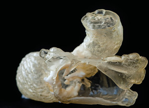
Printed heart: based on magnetic resonance imaging, a portion of the heart showing areas of calcification, in white inside the heartléo ramos
Technological improvements in 3D printers, capable of printing all kinds of plastic parts with polymers, also allows the creation of customized medical models of areas of a patient’s body about to undergo surgery. BioArchitects, a São Paulo-based medical technology company that has been producing biomodels with 3D printers since late 2015, has partnered with a number of surgeons, who are also university professors, to offer the models for testing. In exchange, the surgeons have agreed to report their experiences in working with the 3D replicas in scientific articles. “Based on images from magnetic resonance imaging or tomography, our bioengineers transform these images into three-dimensional models that provide a picture of the patient’s condition,” says Felipe Marques, the company’s chief executive. Producing the biomodel with the shape, texture, flexibility and consistency nearly identical to the real one is made possible by an Israeli-made printer that can use nine different types of polymers in a single impression, mixing some of each, to form skin, subcutaneous tissues and bones. The colors that differentiate organs, tissues and bones can also be chosen with the mixture of polymers.
In Curitiba, south of São Paulo, the company’s biomodel was instrumental in planning a liver transplant surgery in which part of a baby’s abdominal cavity was reproduced on the printer. The 10-month-old child had been diagnosed with biliary atresia, a disease of the bile ducts that only affects infants. To better prepare for the surgery in which a portion of the father’s liver would be implanted in the baby, the doctor sent the baby’s tomography images to BioArchitects in São Paulo. After five days, the material was ready and sent to Curitiba. “In this type of surgery, the right size is very important because an organ that swells too much may lead to compression syndrome in which the abdomen has to stay open until the edema decreases,” notes surgeon Júlio Cesar Wiederkehr, a professor in the Surgery Department of the Federal University of Paraná (UFPR), who faced this situation in early 2016. In addition to this operation, he performed another to remove a tumor from a patient’s liver. “We are often faced with anatomical variations at the time of surgery; the most important thing we need to know is the position of the tumor in relation to the structures of the organ itself as well as to the adjacent ones,” he says. “The use of 3D models may have a number of applications in neurosurgery, orthopedics, cardiac surgery, hepatobiliary surgery, among others,” says Dr. Wiederkehr.
Generally speaking, the use of these customized 3D medical models is indicated for more complex surgeries, when prior planning is required. “Surgeries for congenital cardiopathies and those requiring more than one technique will be better planned if we have a biomodel to simulate the most appropriate correction,” says cardiologist Luiz Antônio Rivetti, a professor at the School of Medical Sciences of the Santa Casa of São Paulo, who has also used the company’s products. He used the 3D model to visualize a left ventricular aneurysm, a pathology that occurs in 10% to 15% of people who experience myocardial infarctions. “By using the model, we knew precisely the extent of the problem, and were certain of the amount of fibrosis that had to be removed,” says Rivetti. “Three operations were performed, with very good results, in which we were able to select the best technique beforehand.” Dr. Rivetti will present the results of using the biomodels at the September 2016 world congress of the World Society of Cardiothoracic Surgeons in South Africa.
Additional benefits
BioArchitects’ biomodels cost between R$4,000 and R$5,000. Felipe Marques, the company’s CEO, predicts that the price will fall as demand increases. “Biomodels for some surgeries, such as multiple rib fractures, in which the surgeon molds titanium plates based on the patient’s anatomical bone curvature, have been shown to reduce costs and surgery time because of prior planning of the patient’s pathology. In one of these surgeries, the surgeon used the biomodel the day before the surgery to simulate the procedure, and then placed the pre-customized plates in the ribs.” According to Marques, there are benefits all around: the hospital increases the number of surgeries per operating room, the insurer can establish predictability when it comes to the use and amount of material, surgeons can plan and predict possible complications in the operation, and the patient is less susceptible to infection.
The 3D printing market is currently about $4.5 billion worldwide, according to A.T. Kearney, a U.S. consulting firm. By 2020, it is expected to reach $17.2 billion. The health sector represents approximately 15% of this total and is expected to grow by about 25% over the next four years.
The use of medical models was measured in a scientific article published in the June 2016 issue of the journal Surgery. It analyzed 158 scientific articles on the use of 3D products between 2005 and 2015. It was conducted by researchers at the French-based Georges Pompidou Hospital and the University of Paris-Sud. The study indicated that three-dimensional printing is becoming increasingly important in medicine, especially in surgeries, and assessed the advantages and disadvantages of using the models. The procedure has already been tried in 37 countries, such as China, Germany and the United States. Studies indicate that 50% of the models were used in surgeries involving the buccomaxillofacial area and 24.7% were used in orthopedic operations. Of the total, 71.5% of the studies reported use of these models in surgeries. A lower percentage, 9.5%, showed the use of 3D printers to produce customized implants or prostheses, and 6.3%, in the design of molds or facial prostheses such as ears and noses. Among the advantages for surgeons in the operating room, the study identified being more able to visualize malformations and anticipate anatomical difficulties, as well as reducing both surgery time and postoperative complications such as hemorrhages and infections. The disadvantages include extensive preparation time for the model and the need for additional time for preoperative planning, in addition to the additional costs of acquiring the model.
Project
Technical-commercial viability analysis of synthetic biological tissue simulators for training in medical procedures guided by ultrasound (nº 2014/50414-9); Grant Mechanism Innovative Research in Small Businesses Program (PIPE); Principal Investigator Felipe Grillo (Gphantom); Investment R$ 100,875.00.
Scientific articles
MARTELLI, N. et al. Advantages and disadvantages of 3-dimensional printing in surgery: a systematic review. Surgery. Online. January 2016.
COELHO, G. et al. New anatomical simulator for pediatric neuroendoscopic practice. Child’s Nervous System. V. 31, No. 2. February 2015.


