 CIREP/FMRP/USPTwo years ago, during a break between one conference and another at a congress about genomics held at Angra dos Reis, on the coast of Rio de Janeiro, Swiss chemist Kurt Wüthrich made a challenging comment about a protein called prion, the abnormal version of which had been associated with the mad cow disease and forced the British government to sacrifice 150,000 bulls and cows between 1985 and 1996. According to Wüthrich, a world authority in the study of protein structures, who won the Nobel Prize in Chemistry last year, it is not possible to think about treating or curing the diseases caused by the prion before getting to know the structure and the function of the normal version of this protein, then almost entirely unknown. A man of few words, the representative of the Swiss Federal Institute of Technology knew that one of the researchers who was already working in this area was in Brazil, but probably did not expect that she would go so far.
Vilma Regina Martins, of the Ludwig Cancer Research Institute, along with specialists from the Federal University of Rio Grande do Sul (UFRGS), the University of São Paulo (USP) in Ribeirão Preto and the Federal University of Rio de Janeiro (UFRJ), discovered that the normal form of the prion which is short for proteinaceous infectious particle participates in a series of processes that are essential for life. It is linked to the formation and maintenance of the memory, with the growth of nerve cells (neurons), and even with the capacity for overcoming neurological problems like epilepsy, marked by tremors and convulsions of greater or lesser intensity and almost always unexpected. The work of this pharmacist-biochemist is earning prominence, not only for responding to the challenge of the Swiss, but also because of the extent of human epilepsies, which affect some 50 million persons in the world and about 1.5 million just in Brazil.
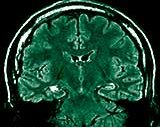
In a recently published article in the Neurology magazine, Vilma’s team and the group from the Epilepsy Surgery Center (Cirep) of the Ribeirão Preto Hospital and Clinics demonstrated that a variant of the gene that encodes the normal or cellular version of the prion, called cellular prion, or PrPc, is a risk factor for the most common type of epilepsy that does not respond to medicines mesial temporal lobe epilepsy. This problem arises as a result of a lesion in a region of the brain called the hippocampus, which participates in important biological processes like learning and the formation of memory. The victims of this kind of epilepsy are treated surgically with the removal of the damaged tissue, a procedure that almost always leads to the cure of the disease. In a group with one hundred patients with this problem treated at the Ribeirão Preto Clinics Hospital, neurologists Roger Walz and Américo Sakamoto, from the Ribeirão Preto Cirep, observed that a small alteration in the gene that produces the normal prion diminished the chances of a cure of the patients after the operation from 92% to 68%. This alteration consists of the replacement of just one amino acid, which is a protein-forming unit: out goes an asparagine, and in goes a serine, and there go the hopes for a good recovery after the surgery.
Vilma and Walz had already shown in mice that the lack of normal prions diminishes the capacity for resisting convulsions, one of the typical, and undesirable, symptoms of epilepsy. In the experiment, they applied the same quantity of three chemical compounds capable of inducing convulsions (kainic acid, pentylenetetrazol or pilocarpine) in two groups of ten mice half of them manufactured the normal prions, and the other half showed, deactivated, the gene that produces this protein. The results showed that the mice without the normal prion are much more sensitive to induced epileptic crises: 90% of them die, due to the convulsions, compared with only 10% of the normal ones, according to the article published in the Epilepsia [Epilepsy] magazine in 1999.
The mice showed a marked difference in another area as well, the memory, according to an experiment carried out in partnership with neurobiologist Iván Izquierdo, from UFRGS, and published in March in Neuroreport. First, the animals are placed on a platform inside a cage. As they like to explore a lot, they come down from the platform and put their four paws on the metal grid at the bottom of the cage: that is when they are given a small electric shock. On the following day, the same animals go back into the cage, but they remain on the platform for a time that is roughly ten times longer, after learning that they will get a shock if they go down to the grid. In fact, the time they remain on the platform in this second stage is used to measure the animal’s capacity for learning. The result was that there was a difference in learning between the two groups one that manufactured normal prions, and the other that produced this protein deactivated. But this disparity only appeared among the adult animals, in their ninth month of life, equivalent to 50 years in the human being. When they are still young, with only three months, which corresponds to almost 20 years in people, there were no differences in learning between the two groups.
With these findings, Vilma proves a relationship between the normal prion a protein that is abundant on the surface of the neurons of a broad group of animals, from reptiles to mammals and the workings of the brain. Discovered at the end of the 1970s by the American researcher Stanley Prusiner, this protein broke one of science’s paradigms, according to which only organisms with genetic material (DNA or RNA), like viruses, bacteria, fungus or worms, could cause diseases the discovery of this protein earned him the Nobel Prize in Medicine for 1997. It was also Prusiner who identified the two versions of this protein: one abnormal and the other, normal, differentiated only by the format: cellular prion shows a helicoidal shape with three stretched out spirals, like the spiral binding of a notebook, united by flexible structures of no defined shape, while in the infectious one of the spirals and the flexible structures form a plane, like a fan.
As a consequence of the format that they take on, the functions of one and the other change a lot. While the normal version of this protein is essential for the proper functioning of the brain, the abnormal one causes the mad cow disease or bovine spongiform encephalopathy, scrapie in sheep, and Creutzfeldt-Jakob disease in human beings. They are rare diseases and up until now incurable, which leave the brain full of holes, with the appearance of a sponge. And it is not just in the brain that the altered prions pile up. In an article published in the New England Journal of Medicine, of November 6, the team led by Adriano Aguzzi, from the University of Zürich Hospital, in Switzerland, found that the altered form of the prions also appears in the spleen and the muscles of the body a finding that may lead to simpler and safer techniques for identifying the Creutzfeldt-Jakob disease. At the moment, diagnosis is based only on the symptoms and on the analysis of nuclear magnetic resonance images of the brain, which does not exclude other diseases of the nervous system, like Alzheimer’s disease. Today, the conformation of the diagnosis is only possible after the death of the patient and an analysis of the brain tissue.
It was also only recently that a possibility of treatment has been arrived at. British researchers injected a chemical compound called pentosan polysulfate directly into the brain of a 19-year old youth in Belfast, Northern Ireland, and succeeded in halting the evolution of the Creutzfeldt-Jakob disease. Luckily, some of these diseases can be contained as their propagation is controlled, which happens mainly through the ingestion of byproducts of contaminated beef and by surgical procedures like the transplant of individuals with Creutzfeldt-Jakob and the use of infected electrodes, which transmit the abnormal protein.
PAGES 24 and 25 FinanceX-ray of innovationInnovative potential of universities and research üthrich’s observation in Angra dos Reis gains consistency to the extent that hypotheses about the origin of the mad cow disease and other similar diseases that attack the brain: it is believed nowadays that the infectious prion is capable of converting the cellular prion into an abnormal prion. This possibility gained strength when biologist Susan Lindquist, a director of the Whitehead Institute, in Cambridge, United States, published an article in the Science magazine demonstrating that this contagion really does occur, at least in mice. Another study, published in Nature of October 16 by the team of Surachai Supattapone, from the Dartmouth Medical School, also in the United States, clarifies a bit more this conversion mechanism of the prion, by suggesting that a molecule of RNA can speed up the transformation of the normal prion into an infectious one.
If the abnormal prion destroys the nerve cells, the normal one has the opposite effect: it prevents the death of neurons. That was what Vilma discovered, this time in a partnership with neurologist Rafael Linden, from UFRJ. Last year, according to two articles published in the magazine European Molecular Biology Organization, the Embo Journal, the two teams found that normal prions protect the nerve cells of the retina of mice from the so-called programmed cell death, also known as apoptosis. “The normal prion may be involved as well in the process of the development of the fetus and in the correct formation of the tissues”, Vilma comments. Normal prions have proved to be equally essential in the process of the growth of nerve cells, particularly when surrounded by a protein called laminin, which helps to join one cell to another. With this protein nearby, the axons and dendrites extensions of the nerve cells produced by neurons of mice embryos grew in 24 hours to about three times their original size in half of the cells. The same result was observed in only 20% of the cells of embryos in which the gene of the cellular prion was turned off. The results were published in 2000 and, according to Vilma, they show that “normal prions may be related to the capacity of the neurons for making new connections between themselves or for reestablishing connections”.
Laminin itself seems to have an important role in the formation of the memory. In a similar experiment, the researchers from the Ludwig Institute and from UFRGS injected drugs that block the action of the normal prion and of the laminin directly into the hippocampus of 200 rats (another 200 served as a control group and were not given anything). The result: the animals from the first group completely lost their memory, incapable both of remembering something they had learnt two hours before, the so-called short term memory, and what they had been taught 24 hours before, the long term memory. “The results (found by Vilma’s group) are of considerable interest”, Wüthrich commented to Pesquisa FAPESP . The Brazilian team, which over the last four years has published 20 scientific articles with partial results in high impact magazines, is now beginning to study the function of the normal prions in cancer. According to the preliminary results, there seems to be a larger quantity of this kind of molecule in tumor tissues of people with certain types of breast cancer. Should the analysis of the data confirm the first impression, measuring the quantity of cellular prions could in future be used in diagnosing some types of cancer and for assessing the evolution of the disease.
Carried out in laboratories with scientific rigor, the studies about the prion point back, oddly enough, to an ancient habit of the inhabitants of the island of Papua New Guinea, in the Pacific Ocean: cannibalism. The Fore, as the people there are called, were the last to quit the habit of eating human legs and brains. They only stopped, 40 years ago, because of an indigestible consequence: kuru, an illness that would begin with shivers, then immobilize the muscles of the whole body, and finally kill the victims with their throats hardened. The women thought that it was a spell cast by the men of the tribe. It was no spell, but the abnormal prion in action.
The Project
Role of Cellular Prion Protein in Physiological and Pathological Processes (nº 99/07124-8); Modality Thematic Project; Coordinator Vilma Regina Martins Ludwig Cancer Research Institute; Investment R$ 2,728,466.87