The capuchin monkeys are some of the most intelligent primates of the Americas. As they age, their brains can present the same types of lesions that characterize Alzheimer’s disease. Researchers from the University of São Paulo School of Medicine (FM-USP) and the University of Brasília’s (UnB) Primatology Center identified in two elderly animals—one 29 and the other 33 years of age—both beta-amyloid peptide plaques, formed by the buildup of these protein fragments around the neurons, and tau protein neurofibrillary tangles, which accumulate inside these cells and kill them. Neither plaques nor tangles were found in the brain of a nine-year-old young-adult animal. The findings were published in the journal Scientific Reports in March, and according to the authors, the findings may pave the way for capuchin monkeys to be adopted as a natural model to study the evolution and treatment of Alzheimer’s disease, the most commonly occurring form of dementia in humans.
The idea of examining whether something similar to Alzheimer’s disease also occurs in capuchin monkeys arose years ago, when the neurologist Ricardo Nitrini, FM-USP, a scholar of dementia epidemiology in Brazil and one of the coordinators of this research, met psychologist Maria Clotilde Tavares. A neuroscience and behavioral specialist, Tavares was at that time the coordinator of the UnB Primatology Center, home to the animals used in the study. “We awaited the natural death of these animals before evaluating the brain tissue,” reports Nitrini.
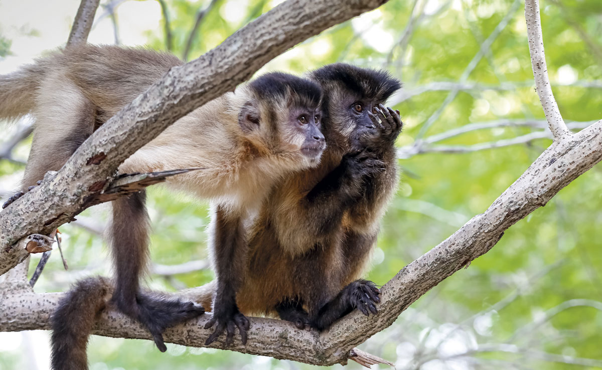
With sizes varying from 35 to 48 centimeters (tail not measured) and weighing up to 4.8 kilograms, capuchin monkeys (Sapajus libidinosus) are among the most common nonhuman primates in the Americas. In Brazil, these monkeys live in practically all biomes, from the most humid, such as Amazonia and the Atlantic Forest, to the driest, such as the Cerrado (wooded savanna) and the Caatinga (semiarid scrublands). The monkeys attracted the interest of those investigating cognitive decline because, in addition to being abundant, they have more sophisticated manual skills and behaviors than other monkeys do, such as the fabrication and use of tools to hunt or extract chestnuts. Furthermore, the encephalization quotient of these monkeys—that is, the size of their brain in relation to their body, an indicator of intelligence—is superior to that of most other nonhuman primates, and their brains are anatomically more similar to those of humans, with folds and grooves, than other monkey species, particularly those native to the continent.
“These animals have significant behavioral plasticity and adapt to different environments. I believe that, with this study, they will be more valued in the investigation of neurological illnesses,” says UnB’s Tavares. “Capuchin monkeys have a long life and can live to 40 in captivity. This will allow study into how brain degeneration affects the skills of these animals over time,” says Tiago Falótico, researcher and current president of the NGO Neotropical Primates Research Group (NeoPReGo). Falótico is examining the use of tools and cultural evolution of capuchin monkeys and was not part of the study published in Scientific Reports, conducted with FAPESP funding, of the Brazilian National Council for Scientific and Technological Development (CNPq), Alzheimer’s Association, and National Health Institutes (NIH) of the United States.
Until recently, neurologists and neuroscientists conjectured that Alzheimer’s disease was an exclusively human disease because there was no knowledge of other animals presenting the two typical types of lesions. This belief began to change in 2008, when Lary Walker and his team at Emory University in the U.S. reported, in an article published in The Journal of Comparative Neurology, the finding of beta-amyloid peptide plaques and tau protein tangles in the brain of a 41-year-old female chimpanzee.
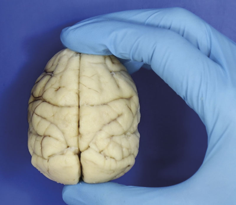
Roberta Diehl Rodriguez / USPHigh encephalization quotient: capuchin monkeys’ brains (real image) are proportionally large in relation to their bodiesRoberta Diehl Rodriguez / USP
Originating in Africa, these simians are, in evolutionary terms, the closest living primates to human beings, sharing a common ancestry from between 7 and 5 million years ago, with almost 99% identical genes. For this reason, from a biological standpoint, chimpanzees appear to be a good model for investigating the evolution and treatment of illnesses afflicting Homo sapiens, including Alzheimer’s disease. However, maintaining a chimpanzee in the laboratory is expensive—according to some estimates, it can cost over US$20,000 per year—and these animals are at risk of extinction (there are fewer than 300,000 in nature). Furthermore, little more than a decade ago, animal research directives became stricter. U.S. and European healthcare authorities have recommended that chimpanzees be used only to investigate human illnesses when there is no other model available or when it is not possible for ethical reasons to carry out these tests on people.
Given the impossibility of conducting certain experiments on human beings, researchers have advanced their understanding of the phenomena behind Alzheimer’s disease and the quest for more effective potential treatments on the basis of initial studies with models that are not always ideal. The experiments generally begin with cell culture in the laboratory, enabling investigation of the gene activation pattern and modifications in the biochemical pathways occurring during disease, and progress to tests on rodents, almost always mice, which can indicate how the disease affects certain cognitive aspects. The variety of models used to understand how the disease takes hold and evolves is comprehensive, ranging from worms to fruit flies and from fish to other primates, such as lemurs and some monkey species. However, none of these models faithfully and completely reproduce what happens in the human brain.
Invertebrates, for example, are advantageous in certain situations because they share certain genes relevant to Alzheimer’s disease and reproduce more quickly than mammals do. However, although invertebrates can be useful for learning about the biochemical pathways affected by disease, they are not ideal for studying treatments because the effects that the compounds produce in them are not always observed in animals with a more complex nervous system.
Rodents are by far the animals most commonly used in research, allowing the observation of certain behavioral effects of the disease, such as the loss of spatial memory, and certain benefits of potential therapies. However, rodent models do not enable full reproduction of the disease. “Mice and rats do not spontaneously develop beta-amyloid peptide plaques, nor tau protein tangles, although the first type of lesion occurs naturally in elderly specimens of a rodent known as the degus, found in Chile,” says the biochemist Sergio Teixeira Ferreira of the Federal University of Rio de Janeiro (UFRJ), who investigates the causes of Alzheimer’s disease.
For at least two decades, researchers have tried to address this limitation by artificially manipulating the animals. One of the strategies is to genetically alter rodents to produce beta-amyloid plaques or tau tangles. Another, developed by the UFRJ group, involves the injection of beta-amyloid peptide fragments (oligomers) directly into the mouse’s brain. “This causes alterations very similar to those brought about by Alzheimer’s disease, including memory loss,” explains Ferreira.
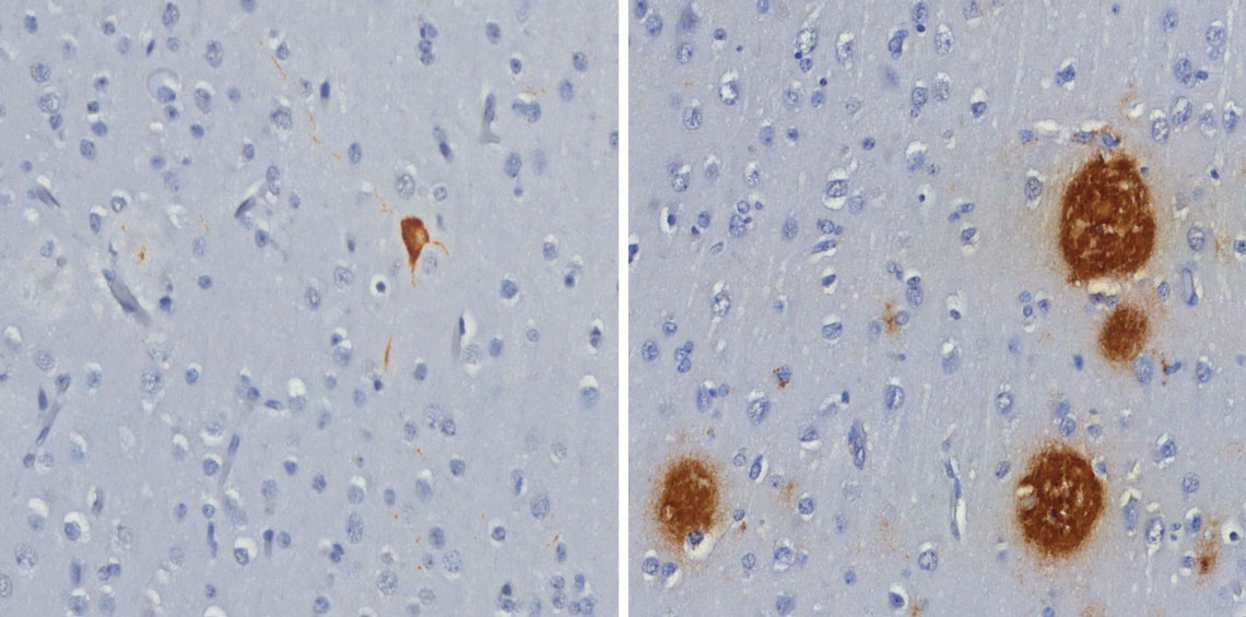
Roberta Diehl Rodriguez / USPTau protein tangle (left) and beta-amyloid peptide plaques (right), both brown, found in the brains of capuchin monkeysRoberta Diehl Rodriguez / USP
Even so, what happens with rodents can be very different from what happens in the brains of humans. For example, mice share only 85% of their genes with humans and have a much simpler brain than humans from an anatomical perspective. In rodents, the brain has a higher ratio of neurons than other cells, whereas in the human brain, the ratio is approximately one-to-one. Researchers estimate that this difference can make the disease develop in animals differently from the way it evolves in humans.
“Despite the success, the two-dimensional models of cells and animal models can capture only a fraction of the mechanisms of Alzheimer’s disease because they are incapable of recapitulating the specific structure, function, and cell diversity of the human brain,” wrote bioengineer Donghui Zhu and colleagues of Stony Brook University, in the U.S., in a 2022 review article published in the journal Bioengineering and Translational Medicine.
One way to close the gap between human disease and animal models is to use phylogenetically closer species that spontaneously develop the typical pathology of Alzheimer’s disease. “Working with natural models allows observation of a more realistic condition, more similar to what happens in humans,” says Analía Arévalo, a neuroscientist specializing in language, and a researcher at the Experimental Surgery Research Laboratory at FM-USP, who did not participate in the Scientific Reports study.
Even among primates, which have more complex cerebral architecture than rodents do and are evolutionarily closer to humans, the perfect model has yet to be identified. Beta-amyloid peptide plaques have been detected in rhesus monkeys (Macaca mulatta), cynomolgus macaques (Macaca fascicularis), white-tufted marmosets (Callithrix jacchus), and gray mouse lemurs (Microcebus murinus), but these plaques do not always occur simultaneously with tau protein tangles, although the animals may present cognitive deficiencies. Another difference is observed in the brain regions where these plaques and tangles form. In humans, plaques and tangles occur more frequently in the hippocampus, the area associated with memory acquisition and consolidation, whereas in marmosets and rhesus monkeys, these structures are more common in areas associated with emotions (limbic system) or hearing (temporal cortex).
In these two aspects, capuchin monkeys may provide a closer model of human disease: the monkeys present beta amyloid plaques and tau protein tangles, and these lesions affect both the cortex and the hippocampus, with researchers also identifying signs of neuroinflammation, which also occurs in humans. “No other New-World primate used in the studies is as intelligent as the capuchin monkey, which makes its own tools and can remain in a biped position for quite some time,” says neuroscientist Roberta Diehl Rodriguez, lead author of the Scientific Reports study. “Moreover, the New-World primate presents more similar neuropathological alterations to those of human beings,” she states.
For capuchin monkeys to effectively become model animals for Alzheimer’s disease, however, researchers need to confirm the occurrence of lesions in a greater number of animals and characterize how these lesions affect behavior. “This will be the most interesting aspect to compare with the disease profile in humans,” says Arévalo.
Even if it works, there is a limitation. The disease in these animals may take decades to develop. To overcome this issue, the neuroscientist Fernanda De Felice’s group at the UFRJ is seeking an artificial Alzheimer’s disease model in young primates. Ten years ago, she and colleagues from Queen’s University in Canada successfully reproduced in cynomolgus macaques—currently endangered—the damage that Alzheimer’s disease causes in humans through the injection of beta-amyloid oligomers into the animals’ brains. These oligomers accumulate in the frontal cortex, the hippocampus, and other areas associated with memory and cognitive aspects. More recently, the group induced lesions in young rhesus monkeys between 3 and 5 years of age to avoid waiting for the natural evolution of Alzheimer’s disease. “If we can reproduce the disease in young animals,” says Ferreira, of UFRJ, and husband and collaborator of De Felice, “it would make the work much easier.”
Published in July 2024
Project
Characterization of tau astrogliopathy in aging and neurodegenerative diseases (nº 16/24326-0); Grant Mechanism Postdoctoral Fellowship; Supervisor Ricardo Nitrini (USP); Beneficiary Roberta Diehl Rodriguez; Investment R$301,733.62.
Scientific articles
RODRIGUEZ, R. D. et al. Bearded capuchin monkeys as a model for Alzheimer’s disease. Scientific Reports. mar. 15, 2024.
ROSEN, R. F. et al. Tauopathy with paired helical filaments in an aged chimpanzee. The Journal of Comparative Neurology. may 14, 2008.
SREENIVASAMURTHY, S. et al. Current progress of cerebral organoids for modeling Alzheimer’s disease origins and mechanisms. Bioengineering and Translational Medicine. jul 29, 2022.