
 reproduction of the painting ‘Everything is false and useless IV’, by Iberê Camargo. 1992Early discovery of changes in the brain that may indicate the onset of Alzheimer’s disease, the main cause of dementia in the elderly, is one of the challenges of the neurology of ageing. Researchers from the School of Medicine of the University of São Paulo (FMUSP) believe they have identified the first region in the brain to show the lesions that are most characteristic of the disease, the so-called neurofibrillar bundles. Alzheimer’s begins in the brain stem, more specifically in an area called the dorsal raphe nucleus and not in the cortex, which is the center for processing information and storing memory, as medicine traditionally claims. This idea is defended by Brazilian scientists in partnership with colleagues from three German universities, in an article to be published over the next few days in the scientific journal, Neuropathology and Applied Neurobiology.
The work’s conclusion is based on the autopsy of the brains of 118 people whose average age was 75 at the time of their death. Researchers noticed the existence of lesions in the dorsal raphe nucleus in 8 of the old people that had no bundles in other parts of the brain and in all the 80 individuals who already had at least one bundle in the transentorhinal cortex, the region classically indicated as the first to be affected by Alzheimer’s. The 88 individuals with anatomical marks on their brains associated with this type of dementia showed varying degrees of clinical manifestation of the disease and some might even have been asymptomatic. The work of the Brazilians and Europeans relied on multiple sources of funding: money form German institutions, the National Council for Scientific and Technological Development (CNPq), the Albert Einstein Israeli Teaching and Research Institute in São Paulo, and FAPESP, which is funding a line of studies by neurologist Ricardo Nitrini, from FMUSP, into the incidence of dementia in the Brazilian population.
The stem, which is responsible for connecting the cortex to the spinal medulla, is not strictly part of the brain but of the encephalon that includes the brain, the cerebellum and the stem. Put simply and resorting to metonymy, a figure of speech in which part can be used for talking about the whole, lay people use the word brain almost as a synonym of encephalon, although technically speaking that is not what it is. Technical definitions aside, the brain stem is an important structure of the nervous system: it controls involuntary functions that are crucial for survival, like breathing, heart beat, blood pressure, sleep and even dreams.
NIH
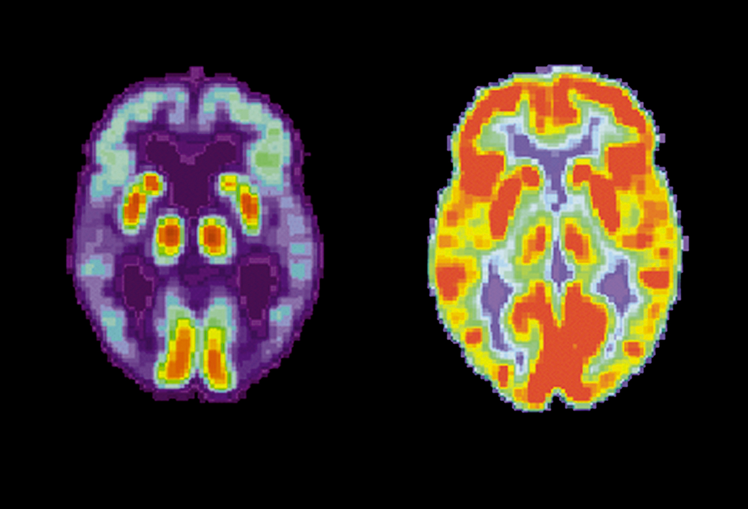
Tomography of a brain with Alzheimer’s (left) and another normal oneNIHIf the discovery that Alzheimer’s disease starts in the brain stem, the smallest of three large parts of the encephalon, and from there spreads to interconnected areas of the cortex, is confirmed by new studies, this is an important piece of information in the search for therapies to slow the development of the disease in its initial stage. This fact may mean that this region of the nervous system becomes the preferred target for actions relating to new drugs and therapies against the disease. “We need to know where the initial lesions appear in order to try and discover efficient forms of delaying the development of Alzheimer’s when it is still in its very early stages”, says pathologist Lea Grinberg, coordinator of FMUSP’s human brain bank, the source of brain samples for the study and the primary author of the article. “Our study confirms that the brain stem is obviously the first region vulnerable to Alzheimer’s and the point from which this devastating disease spreads”, says Helmut Heinsen, from the Psychiatry Institute of the University of Würzburg, one of the German researchers who shares authorship of the work with the Brazilians. “We hope that the results we’ve obtained with human models give new impetus to the development of prevention and treatment strategies for Alzheimer’s.” Injections of stem cells, transplants of reprogrammed brain cells, image-guided immunotherapy – all these still-evolving techniques may one day be tested on the dorsal raphe nucleus as candidates for therapy.
The neurofribillar bundles appear due to a chemical alteration in the structure of the tau protein responsible for the formation of the microtubules that transport nutrients and information from the neuron prolongations to its cell body and vice-versa. When modified the protein destabilizes the microtubules, leading to a collapse of this system and the death of neurons. Along with the appearance of extra-cellular plates arising from the abnormal accumulation of the protein, Amyloid beta, a second type of lesion also closely associated with the occurrence of Alzheimer’s, the presence of the bundles is the trademark sign of the progression of the disease in the brain. There is no agreement among specialists about which of the anatomical changes, the bundles or the plates, is more important for the development of this form of dementia. “But there’s evidence that the progression of the bundles is more crucial than that of the Amyloid beta protein plates to determine the clinical seriousness of Alzheimer’s”, adds Lea.

nihTwo types of lesion associated with Alzheimer’s: neurofibrillar bundles…nih
Forgotten region
Confirmation that the dorsal raphe nucleus has neurofibrillar bundles in Alzheimer’s is nothing new. Science believed, however, that the lesions at this point in the brain stem came after and not before sectors of the cortex had been affected by the alterations that are typical of the disease. In fact, not much importance was given to this part of the brain in pathological examinations looking for anatomical alterations associated with dementia. “Until this new work by the Brazilians and Germans no one who worked with Alzheimer’s looked at the brain stem”, comments biochemist Sergio Teixeira Ferreira, from the Federal University of Rio de Janeiro (UFRJ), who is studying the disease. “The great merit of this study is that it threw light on this region.”
In the so-called Braak and Braak system, which classifies Alzheimer’s lesions into six stages as a function of the brain area taken over by bundles, the whole focus is on the area of the cortex that is closely linked to the question of memory. Degree 1 changes, the lowest on the scale, are those that restrict the transentorhinal cortex, the point in the brain usually described as being where Alzheimer’s starts. With this system no account is taken of the existence of lesions in the brain stem (or even in the dorsal raphe nucleus, which is more or less three centimeters from the transentorhinal cortex) to attribute the possible degree of the extent of Alzheimer’s. “We’re suggesting including lesions in this region of the brain stem as a neuropathological stage prior to the present stage 1 of the Braak and Braak system”, write the authors of the scientific article in Neuropathology and Applied Neurobiology. “The finding [of the article] really is something new in its field and much better founded”, comments neuroscientist Ivan Izquierdo, from the Pontifical Catholic University of Rio Grande do Sul (PUC-RS), one of the world’s authorities in the study of the mechanisms of the formation and extinction of memory. “The authors of the study are to be congratulated.”
Alzheimer’s, a neurodegenerative disease, the origin of which is still a mystery, that still has no cure and the brain lesions of which lead to the progressive death of neurons, to growing memory loss and subsequently the loss of other cognitive functions to the point of compromising the carrying out of trivial tasks, like crossing the street, recognizing a relative or brushing teeth, affects mainly those over 60. The ageing of the world’s population (a tendency also seen in Brazil, where the number of elderly, today around 19 million people, is likely to double over the next 20 years) means that the disease is currently seen as one of the priorities for medical research. It is difficult to fight Alzheimer’s, given that its progression may be silent. From the moment the initial brain lesions arise to the appearance of the first clinical symptoms of cognition loss, more than a decade may have passed. There is only one way of unmistakably diagnosing the disease: carrying out an autopsy of the brain to look for the anatomical changes that are typical of the disease. Clinically, before carrying out the autopsy, it is impossible to be 100% certain that an elderly person is suffering from Alzheimer’s, above all if he/she is at the start of the cognitive loss process. In short, not everyone who forgets things, whether they are elderly or not, is necessarily suffering from Alzheimer’s or some other type of dementia.

nih…and Amyloid beta protein platesnih
Seeking a cure for Alzheimer’s disease is a very valid ambition for medical research. But in the short term perhaps it is more realistic to think about ways of slowing down the progression of the brain lesions that lead to Alzheimer’s and delaying as far as is possible the appearance of cognitive problems that little by little reduce the patient’s quality of life. In this way medicine would reduce the morbidity period of the disease. “If we manage to delay the appearance of the clinical symptoms of Alzheimer’s disease by ten years this would be the equivalent of practically eliminating the disease in the elderly”, explains neurologist Ricardo Nitrini, from FMUSP, another of the study’s authors.
Unique in Brazil FMUSP’s brain bank began being established in 2004, basically with resources from the School of Medicine itself, the Ministry of Health and the Albert Einstein Israeli Teaching and Research Institute. Today it has a collection with almost 2000 samples of nerve tissue from people who were at least 50 years old when they died of natural causes and, as a result, an autopsy was carried out by the Death Verification Service (SVO) of São Paulo, which is linked to FMUSP. Annually the SVO carries out some 13,000 autopsies. Only the brains of dead people whose families have agreed to donate them for research are used in scientific studies. In addition to authorizing use of the encephalons the relatives have to consent to respond to questions aimed at checking if the dead relative had any cognition loss or clinical manifestation that might be associated with some aspect of dementia. “The willing reception of people to our studies was surprising”, comments Lea, who spends most of the year in Germany where she is doing post-doctorate studies at the University of Würzburg, with a grant from the Humboldt Foundation. When they have the data from family members the researchers crosscheck the clinical diagnosis of the donor with the results of the histo-pathological examination of his/her brain. In this way they arrive at a final verdict about the donor’s condition: if they were normal or had some form of dementia, with just clinical symptoms or also with anatomical lesions.
As the encephalon collection that forms part of FMUSP’s Brain Ageing Project grew and became better structured it started supplying nerve tissue samples to various research groups from the university itself and from other institutions. One of the first pieces of work done with this material is precisely what can be discovered from where the Alzheimer’s lesions appear in the human brain. But there are other studies on-going, some with already tangible results and others still in their initial stages. The DNA of brains was extracted and sent to the Center for Human Genome Studies at USP, which is going to try and find genes linked to the occurrence of dementia. Helena Brentani, from the A.C. Camargo Cancer Hospital is studying the expression of the RNA molecule in the brains of patients with Alzheimer’s. In another line of research aimed at evaluating brain alteration arising from ageing, Roberto Lent’s team from UFRJ carried out interesting work with samples supplied by USP. “The brain bank is exemplary and very well organized”, says Lent. With the help of their own methodology researchers from Rio estimated the number of neurons and other types of cells in brains taken from healthy individuals from the cognitive point of view. Some of the results are surprising.
 Most common dementia
With a rich sample of nerve tissue readily available the researchers of the Brain Ageing Project have the chance to confirm, now with the help of modern and accurate examinations, epidemiological information about the incidence of dementia in the Brazilian population. Because they have a very large collection of brains representing so many individuals who were healthy in terms of cognitive disease as well people who had various types of neurological problems, they can design studies with highly varied end purposes. Preliminary data from this work confirm that Alzheimer’s is really the most common cognitive disease among the elderly, corresponding to almost 60% of the cases, a figure similar to that normally put forward by clinical studies. This was followed by vascular diseases (25%), Lewy’s corpuscle disease (10%) and other cognitive disorders. “These other dementias are generally under-diagnosed from the clinical point of view”, explains Lea.
Hypertension and diabetes are diseases associated fundamentally with heart problems. But they should also be more frequently seen as risk factors for dementia. In a sample of brains that were somewhat compromised cognitively the group from USP noticed the existence of microvascular alterations in half of them, a high figure. “These alterations have a direct impact on cognition becoming worse, whether the person has Alzheimer’s or not”, comments Nitrini. Another intriguing fact that arises from these studies is that some 40 people over 80, who from the neuropathological point of view had typical Alzheimer’s lesions in their brains, had no clinical manifestations of dementia, according to members of their families. This may indicate that these individuals had something that neutralized the damaging effects of the lesions, perhaps some protection factor, with a probable implication on the treatment of the disease. “We’re trying to understand why these people did not get sick”, explains Wilson Jacob-Filho, a professor of geriatrics at FMUSP and general manager of the Physiopathology in Ageing Laboratory. “This may have occurred due to some genetic characteristic or behavioral and environmental determinants during their lives.” As can be seen there is no lack of mystery about Alzheimer’s not yet unveiled.

Nicolas RougierDrawing of a neuronNicolas Rougier
The geography of nerve cells
Three out of every four human neurons are in the cerebellum and not in the cortex, says the work by the Brazilians
Of all the animals the brain of humans is the most complex because it has more neurons in the cortex, right? Neuroscientists Suzana Herculano Houzel and Roberto Lent, from the Federal University of Rio de Janeiro (UFRJ), who have just finished counting neurons in human brain samples supplied by the brain bank of the University of São Paulo (USP), do not agree with this dogma of science. For them, who should shortly publish a study on the subject, the major difference with Homo sapiens lies in the number of nerve cells in the cerebellum. This part of the brain that is responsible for maintaining balance and motor coordination represents a little more than 10% of the size of the encephalon, but according to the calculations of the researchers, houses approximately 75% of all human neurons. “The cortex has no more than 20% of our neurons”, says Lent. Nevertheless, its weight is the equivalent of 70% of the encephalon. “Looked at in this way the cerebellum and not the cortex represents the pinnacle of human evolution.” The rest of the human neurons are distributed in smaller structures, like the bulb.
For the neuroscientists the discovery was not totally unexpected. On the contrary, the pair from UFRJ, who have developed their own method for counting neurons, have published scientific work over the last three years showing that in 13 different species of mammal (six rodents, six primates and a tree shrew) the number of neurons in the cerebellum increases as a function of brain size, while the number of nerve cells in the cortex varies proportionally much less relative to the dimension of the encephalon. In rodents the cortex had on average only 18% of all the neurons and most of the nerve cells were in the cerebellum. In non-human primates the percentage of neurons in the cortex varied between 19% (bush-baby) and 42% (squirrel monkey) of the total, indices always smaller than those found in the cerebellum.
According to the work of the researchers the total number of neurons in the human brain is not much different from what is normally suggested. They counted some 90 billion nerve cells. The classic number says that the encephalon of our species houses some 100 billion neurons. The small difference is perhaps due to the fact that they counted the neurons of elderly individuals who may have lost some of the nerve cells with the passage of time. If the total number of neurons stays as expected the same cannot be said of the number of glial cells that serve as support and nutrition for the neurons: the team from UFRJ found almost 90 billion glial cells, more or less one for each neuron. “The scientific books say that there are ten glial cells for every neuron”, Lent comments. “But we found far fewer.”
The Project
Nosological diagnosis of dementia in the Brazilian population (06/55318-1); Modality Regular line of research help; Coordinator Ricardo Nitrini – FMUSP; Investment R$ 125,595.45 (FAPESP).
Scientific article
GRINBERG, L.T. et al. The dorsal raphe nucleus shows phospho-tau neurofibrillary changes before the transentorhinal region in AD. A precocious onset? Neuropathology and Applied Neurobiology, to be published on-line.