
Léo Ramos Chaves
Electrode used in the design of devices to detect antigens and antibodiesLéo Ramos ChavesRecent advances in the field of molecular biology are extending the possible uses of biosensors in diagnosing and preventing disease. These devices were designed based on elements of biological recognition such as antigens and antibodies, and could become as portable and cheap as equipment used to measure blood sugar levels. Biosensors are widely used in other countries and are attracting increasing attention from Brazilian research groups, which in recent years have invested in devices designed specifically to detect neglected infectious diseases associated with poverty and the lack of basic sanitation. This is the case with researchers in the Nanomedicine and Nanotoxicology Group at the University of São Paulo São Carlos Institute of Physics (IFSC-USP). Since 2010, this group has worked on developing a set of sensors capable of identifying signs of several diseases. If these prove effective during the next phases of testing, the devices may become an alternative to laboratory exams and could be used in doctors’ offices or by health agents visiting the homes of people who live in remote areas of the country.
In the United States, biosensors have been used for some time by physicians to speed up test results, or to monitor the health conditions of individuals with diseases such as AIDS and hepatitis C. In other situations, they can help measure the levels of oxygen or alcohol in the blood, which is possible using a flexible biosensor created by researchers at the University of California, San Diego. The National Institutes of Health (NIH) has also invested in research to design medical biosensors based on a variety of systems including chemical attraction, electrical currents, and light detection.
At IFSC-USP, one of the medical biosensors which has reached a more advanced stage of development is for diagnosing dengue fever, a disease that affects 390 million people worldwide per year according to the World Health Organization (WHO). The device is based on identification of the NS1 protein, which the virus secretes into the bloodstream in the first few days after infection. This protein, an antigen, induces an immune response in the human body to produce antibodies against it. The problem is that this only happens after the fifth day, which complicates early detection of the disease. To accelerate this process, the physicists Nirton Cristi and Alessandra Figueiredo (under the guidance of materials engineer Valtencir Zucolotto and physicist Francisco Guimarães) have developed a system to diagnose dengue based on IgY immunoglobulin, the antibody that combats NS1.
Léo Ramos Chaves
Quartz crystal microscale…Léo Ramos ChavesThe IgY was isolated from chickens inoculated with NS1, and then immobilized on a gold electrode coupled to a circuit which has a constant flow of electrons. The idea is to conduct the exam using a drop of blood on the device. If the infection is present, when the IgY immunoglobulin comes in contact with the NS1 it will alter the flow of electrons, producing a signal which is recorded and processed by software. The results are ready in 20 minutes or less. “The higher the concentration of NS1 in the electrode, the more intense the change in electrical potential will be,” explains Cristi, who currently is a professor at the Institute of Science and Technology at the Federal University of São Paulo (UNIFESP) in São José dos Campos. The project was developed in conjunction with the company DNApta Biotecnologia of São José do Rio Preto, São Paulo, which shares the patent rights for the technology.
Using a different approach, professor Maria Rita Sierakowski and doctoral student Cleverton Luiz Pirich of the BioPol Group at the Federal University of Paraná (UFPR) created a biodevice that detects NS1 using a quartz microscale with piezoelectric sensors which can generate electrical current when deformed by mechanical pressure. The system was developed in collaboration with the USP Institute of Chemistry. It is composed of a quartz crystal, a gold electrode coated with polyethylenimine, and nanofilms from bacterial cellulose nanocrystals which are modified to react chemically when they come into contact with NS1, changing the frequency and energy-dissipation patterns in the nanocrystals. “In this way, when a serum sample containing NS1 is placed on the biosensor, a software program can be used to check whether the protein is joined to the surface of the material by detecting mechanical microvibrations,” explains Sierakowski.
The biosensor for diagnosing dengue incorporates a number of other devices created by the researchers from São Carlos. All are based on electrochemical systems that alter patterns of electrical signals when they detect specific biological events. One of the first biosensors they designed can identify and distinguish antibodies for leishmaniasis and Chagas disease; today these are diagnosed using the Elisa test, which can detect specific antibodies in blood samples. Despite being widely used in clinical analysis laboratories, the Elisa test cannot differentiate between the antibodies produced against infections caused by these diseases, and consequently requires additional examinations.

Cleverton Pirich
…used by researchers at UFPR in developing equipment to diagnose dengueCleverton PirichThe biodevice was developed in partnership with various Brazilian research institutions. It is composed of electrical circuits printed onto small electrodes; on top of these circuits is placed a set of antigenic proteins isolated from the protozoan Leishmania amazonensis, one of the species that cause tegumentary leishmaniasis in Brazil, or Trypanosoma cruzi, which causes Chagas disease, an illness that affects approximately 8 million people per year worldwide according to the WHO. “If the antibody of interest is present in the analyzed sample, the connection between it and the antigenic protein produces a change in electrical response of the electrode, signaling the presence of the pathogen,” explains Zucolotto. With the IFSC biodevice, which is in the prototype stage, results take about 20 minutes to appear, and a patent for this technology has been filed with the Brazilian National Institute of Industrial Property (INPI).
Genetic analysis
More recently, the team from São Carlos also began working on designing genetic sensors to recognize specific sequences of genetic material from viruses and mutations that are associated with tumors. In 2016, Zucolotto, the physicist Laís Ribovski, and the chemist Bruno Campos Janegitz, from the Center for Agrarian Sciences at the Federal University of São Carlos (UFSCAR), created a sensor capable of identifying the 185delAG mutation, which is associated with breast and ovarian tumors. In their experiment, the group immobilized an excerpt of the region of DNA where this mutation occurs on the surface of the device. When the corresponding sequence is placed on the electrode, the connection triggers a change in the equipment’s electrical response, indicating the presence of the target sequence which is linked to the mutation. The results of tests using this device were described in an article published in the Microchemical Journal. For now, only sequences synthesized in the laboratory have been used.
This same technique was used to differentiate infections caused by the Zika virus and dengue fever. Like dengue, Zika also secretes significant quantities of the NS1 protein into the bloodstream in the first days after infection, which can have a negative impact on analysis using the biosensors which previously were developed for dengue. Using tools from bioinformatics, the group identified specific regions within the genetic material for the two viruses so that each can be secured on the surface of an electrode. The connection between the sequence on the device and the other in the sample produces a change in the electrical signal, indicating the presence of genetic material from the viruses. The sensor has shown good results in preliminary testing involving sequences synthesized in the laboratory.

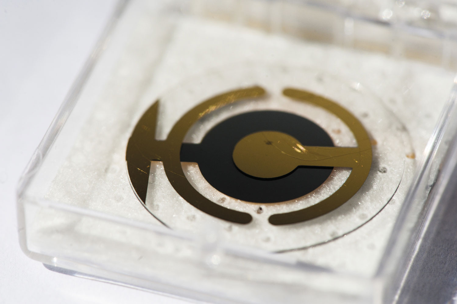
Léo Ramos Chaves
One of the electrodes developed by the group at the São Carlos Institute of PhysicsLéo Ramos ChavesTechnology transfer
Biosensors are growing in popularity around the world. According to data from Markets and Markets, a research and consulting firm in the United States in the area of information technology, the biosensor market was valued at roughly US$16 billion in 2016, and is projected to reach US$27 billion by 2022; medical biosensors account for 66% of this market. The main companies that are investing in the design of this equipment are Abbott Laboratories and Johnson & Johnson (US) and Bayer Healthcare (Germany). Demand for these devices should soar in the coming years, particularly because of the growing prevalence of diseases such as diabetes and the need for devices to monitor blood glucose levels. According to the International Diabetes Federation, this disease will affect 552 million people worldwide in 2030.
The United States is the main market for biosensors, since physicians and researchers in this country tend to rapidly adopt technologically advanced products. In Brazil there is still a long road ahead until these devices are produced and marketed on a large scale. Most of this equipment is developed in universities and public research centers; after being designed in the lab, it still needs to be tested for safety and effectiveness, be patented, and especially attract the interest of companies willing to bring it to the consumer market.
Laboratory-scale production of the electrodes used in biosensors is estimated to cost roughly US$2. “The idea is to develop complete devices with a unit value that does not exceed US$100 (about R$315),” says Zucolotto. Still, most of the devices developed in Brazil are in the prototype stage, and have not been approved by the Brazilian National Health Surveillance Agency (ANVISA). There is also a long way to go until these devices are able to deal with the large volumes of samples that are analyzed in laboratories every day. “In addition to the potential application of this technology in large laboratories, biosensors must be able to analyze, identify, and quantify elements of clinical interest in large numbers of samples,” says physician Jeane Tsutsui, executive director of Fleury Medicina e Saúde. “Before biosensors reach medical clinics and health agents, investment in joint projects between companies and universities is necessary for clinical validation,” she concludes.
Projects
1. Development of electrochemical sensors and biosensors for various analytical purposes (No. 15/19099-2); Grant Mechanism Regular Research Grant; Principal Investigator Bruno Campos Janegitz (UFSCAR); Investment R$169,539.83.
2. Study of the interaction between nanostructured materials and biological systems: Applications to the study of nanotoxicity and development of sensors for diagnosis (No. 08/08639-2); Grant Mechanism Regular Research Grant; Principal Investigator Valtencir Zucolotto (IFSC-USP); Investment R$368,230.90.
Scientific articles
RIBOVSKI, L. A label-free electrochemical DNA sensor to identify breast cancer susceptibility. Microchemical Journal. V. 133, p. 37–42. July 2017.
FIGUEIREDO, A. et al. Electrical detection of dengue biomarker using egg yolk immunoglobulin as the biological recognition element. Scientific Reports. Jan. 2015.
PIRICH, C. L. et al. Piezoelectric immunochip coated with thin films of bacterial cellulose nanocrystals for dengue detection. Biosensors and Bioelectronics. V. 15, No. 92, p. 47–53. June 2015.