When someone is asked to pick up a pencil with their left hand and name the object aloud, information is exchanged between the two hemispheres of the brain. The area responsible for tactile sensitivity in the left hand, located in the right hemisphere, transmits signals to the area in the left hemisphere that processes speech. It has long been known that what makes this communication possible is a structure called the corpus callosum, a robust bundle of tens of millions of white-matter fibers known as axons—extensions of neurons (the nervous system’s executive cells). It functions as a bridge, allowing information to be exchanged between different regions of the two hemispheres. In humans, the corpus callosum can reach up to 10 centimeters (cm) in length and almost 2 cm in thickness.
A study published in the journal Cerebral Cortex by Brazilian researchers in August, however, indicates that the corpus callosum is not the only communication pathway between the right and left sides of the brain. There are other, more subtle routes that remained hidden until they were recently described and mapped by the team. Known as thalamic commissures, these thinner bundles of white matter cross the thalamus, a brain structure located just below the corpus callosum. Approximately 4 cm long and oval-shaped, there is a thalamus in each hemisphere, responsible for processing and relaying sensory information to brain areas that control movement, as well as regulating consciousness, sleep, attention, and memory.
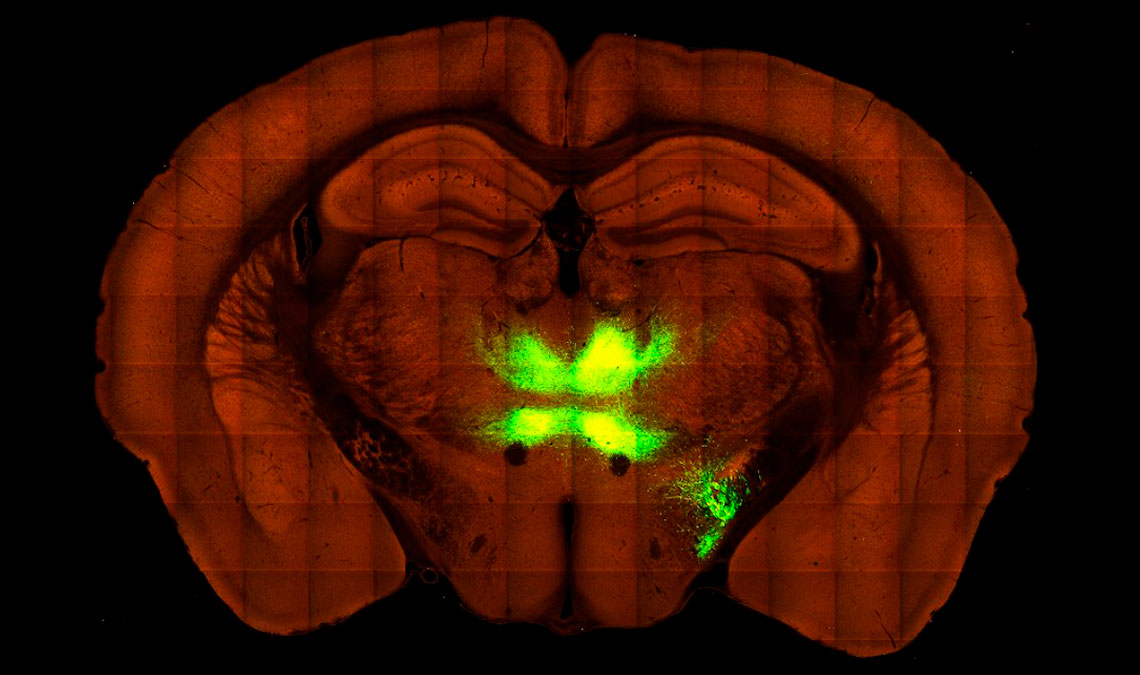
Biomedical scientists Pamela de Meneses Iack and Diego Szczupak began to unravel the mysteries of the thalamic commissures while working at neuroscientist Roberto Lent’s laboratory at the Federal University of Rio de Janeiro (UFRJ). They described the structures in an article published in the journal Cerebral Cortex in 2021. Later, while pursuing a PhD supervised by Lent, Iack began mapping the brain regions connected by these commissures. She injected a genetically modified virus capable of producing fluorescent proteins into different areas of the cortex (the outermost layer of the brain) in rodents and then tracked the path taken by the infectious agent. In parallel, she compared her results with public data from the Allen Mouse Brain Atlas, a 3D map of the connections in mouse brains created by the Allen Institute, which was founded in 2003 by Paul Allen, one of the cofounders of Microsoft. She was thus able to trace the route of the fibers that pass through the thalamic commissures and identify the brain areas connected by them. The research, however, had to be completed by her colleagues, after Iack unexpectedly died in 2024, at the age of 29.
To honor Iack, who conducted the experiments and analyzed the data, Szczupak and the rest of the team completed her work and found a journal willing to accept the biomedical scientist as a posthumous author. The commissures mapped by the group are bundles of axons that originate in the cerebral cortex of one hemisphere and connect to the thalamus of the opposite hemisphere, the researchers showed in the article published in August this year.
It was already known that there are reciprocal connections between the cortex and the thalamus of the same hemisphere. They allow the brain to receive information from the senses and peripheral regions of the nervous system, organize it, and retransmit it to the outer layers of the brain, such as the cortex. But the connections between the cortex and the thalamus of opposing hemispheres, identified by Iack and her colleagues, were a surprise. “These circuits are less dense and therefore not as visible,” says Lent. “That might explain why they had not appeared in the specialized literature before.”
According to Szczupak, now a professor at the University of Pittsburgh in the USA, the existence of these commissures adds a new level of complexity to how the thalamus functions. “The thalamus is a bit like a conductor leading an orchestra,” he explains. “The fact that these fiber bundles cross the hemispheres suggests that it may not only be responsible for doing this within a single hemisphere, but in both.”
The group also found that the commissures are not evenly distributed throughout the central nervous system. Most are located toward the front of the brain, close to the prefrontal cortex, an area associated with decision-making processes. These processes involve handling information from different regions of the nervous system, which may help to explain why there is greater connectivity there. “The thalamus integrates the signals and sends them to the prefrontal cortex as a whole, providing sensory information about what is happening around the individual, which contributes to decision-making,” explains neuroanatomist Newton Canteras of the Institute of Biomedical Sciences at the University of São Paulo (USP).
Although Iack and her colleagues conducted their studies on mice, the same structures have been observed in primates and may exist in humans. Identifying them in people, however, is a more difficult task. “Currently available imaging technologies for the human brain are still rudimentary and do not provide the degree of precision needed to identify individual nerve fibers, as can be done with animals,” explains Lent.
For the UFRJ group, publishing the map of the fibers that make up the thalamic commissures was more than simply an academic achievement. “Knowing that Pamela’s legacy has been read by other people brings some comfort,” says Szczupak. “It is an emotional side of science that does not often appear in the coldness of scientific publications,” Lent concludes.
The story above was published with the title “Complex connections” in issue 357 of November/2025.
Scientific articles
IACK, P. M. et al. Comprehensive mapping of the thalamic commissures in the rodent brain. Cerebral Cortex. Aug. 2025.
SZCZUPAK, D. et al. Direct interhemispheric cortical communication via thalamic commissures: A new white-matter pathway in the rodent brain. Cerebral Cortex. Oct. 2021.