To evaluate the quality of images generated by computed tomography (CT) scanners and define protocols for detecting nodules, radiologists, biomedical scientists, and medical physicists use devices that function as test dummies. Because excessive radiation poses health risks, studies cannot be conducted directly on humans, so researchers place simulator objects inside the machines to calculate the optimal dose for examining specific regions of the body. These objects are known as phantoms.
At the University of São Paulo (USP), a project led by physicist Paulo Roberto Costa, from the Department of Nuclear Physics at the Institute of Physics (IF), has resulted in a new phantom designed specifically for lung CT scans, an exam used for the early detection of one of the world’s deadliest cancers. In 2022, lung cancer caused more than 1.8 million deaths globally. In Brazil, it claimed 28,600 lives in 2020.
“Our phantom combines traditional, well-established features used to optimize CT equipment with the ability to produce images that resemble the human lung,” Costa explains. The device is hybrid: it simultaneously evaluates and quantifies physical image-quality parameters and the anthropomorphic characteristics of the organ, such as the different types of tissue found in the lung. Development began five years ago with support from FAPESP, and the team’s findings were published in August in the journal Medical Physics.
The phantom developed at IF-USP is partially based on a model created at Duke University in the United States, which was later adapted and commercialized under the name Mercury by Sun Nuclear—a device not specific to any human body part. Mercury provides quantitative information such as image resolution, contrast, and noise to test CT scanners. In contrast, the Brazilian team used 3D printing techniques and resins to give their phantom an anthropomorphic design capable of generating realistic tomographic images, similar to those obtained from patients in routine radiological practice. The 3D models were printed at the Renato Archer Information Technology Center in Campinas (SP).
“Commercial phantoms like Mercury are useful for testing CT scanners comprehensively, but they cannot provide a detailed evaluation of the distinctive properties of specific body regions,” Costa explains. For that reason, the São Paulo group chose to focus on the unique characteristics of the lung. According to the physicist, radiological image quality is directly proportional to the radiation dose used—high doses yield better images, while low doses can impair clinical results. “The balance between patient safety and good image quality is difficult to achieve.”
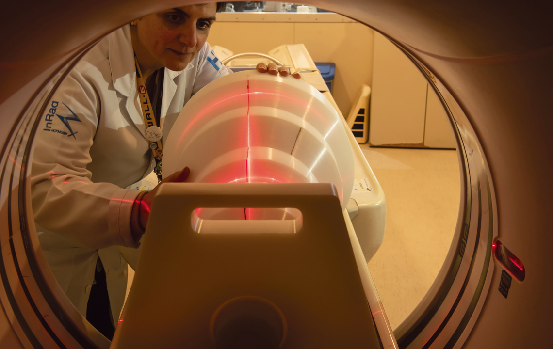
Léo Ramos Chaves / Pesquisa FAPESPCT scan simulation using the phantom developed at USPLéo Ramos Chaves / Pesquisa FAPESP
International collaboration
The project involved collaboration with radiologists specializing in lung imaging, including Marcio Sawamura, director of the clinical staff at the Institute of Radiology at the Hospital das Clínicas of the USP School of Medicine (FM-USP). “I supported the medical part of the project. We selected real patient images to use as models. We chose images of lung nodules and tried to build models that replicated them. Then we reviewed the images produced with different materials used to simulate the nodules to determine which looked most realistic,” Sawamura explains.
Part of the phantom’s validation took place at Radboud University Medical Center in Nijmegen, the Netherlands, between 2023 and 2024, where it underwent measurements on two state-of-the-art CT scanners. “I spent a year in the Netherlands on a FAPESP research grant abroad. During this time, there was extensive collaboration with radiologist Bram Gertus to arrive at a reliable phantom model,” Costa says.
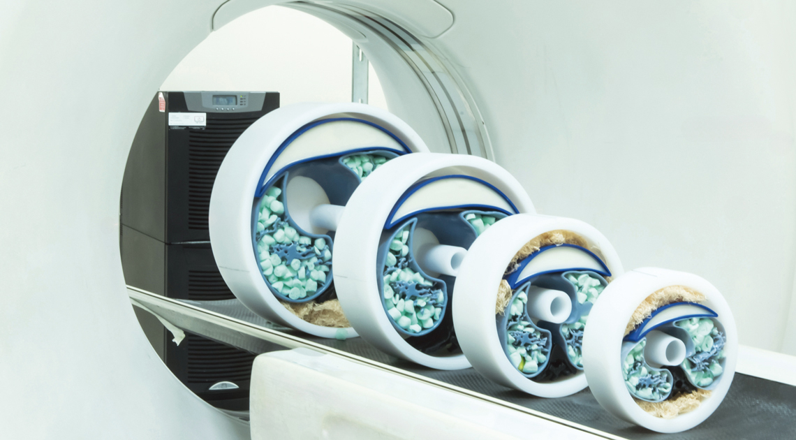
Costa transported the phantom, which weighs approximately 132 pounds, in his luggage. “It has a heavy solid section. When standing upright, it looks like a wedding cake,” he notes. The main structure is made of ultra-high molecular weight polyethylene and includes snap-on modules with five different diameters, from 12 to 37 centimeters, representing patients of various sizes. It is used in two configurations. In the traditional setup, small plates inside the structure capture numerical data describing the CT scanner’s key characteristics. In the anthropomorphic configuration, an alternative set of inserts is placed—like assembling a jigsaw puzzle—to mimic lung tissue properties and various types of nodules.
“I consider it an excellent and forward-thinking project. It is extremely challenging to build an anthropomorphic phantom to simulate the lung because of the complexity of its anatomical structures,” says physicist Diana Rodrigues de Pina, from the Department of Infectious Diseases, Dermatology, Diagnostic Imaging, and Radiotherapy at the Botucatu School of Medicine, São Paulo State University (UNESP), who was not involved in the development.
Pina notes that the USP-developed product will fill an important gap in Brazil. Although lung phantoms for CT scans exist internationally, they are expensive—around R$130,000—and difficult to access. According to the UNESP physicist, a major strength of the Brazilian development is that it is a hybrid device. “It will help radiologists evaluate images and allow national companies that provide CT scanner quality-control services to implement protocols,” she says.
Costa is currently negotiating a partnership with a company to refine the prototype. “What we have now is a proof of concept. Over time, we hope to produce a commercial version,” he explains. “Now that we have demonstrated its applicability and potential for optimizing CT scanners, we’re working on additional tools to improve case studies for cancer diagnosis.” One planned innovation is the development of other anthropomorphic models to simulate pneumonia, infections, and additional findings seen on CT images. The team is also considering the use of AI tools to quantify nodule volumes.
The strong results achieved with the lung phantom motivated Costa to pursue two new projects. “We are refining an abdomen model, made of 3D-printed components representing the liver, spleen, and kidneys, and will soon begin validation tests,” he says. “Another device, designed to simulate the skull, is already at an advanced stage.”
The story above was published with the title “Lung simulator” in issue 357 of November/2025.
Projects
1. Additive manufacturing techniques in the production of computed tomography phantoms (n° 22/11457-0); Grant Mechanism Regular Research Grant; Principal Investigator Paulo Roberto Costa (USP); Investment R$144,921.18.
2. Development, validation, and application of techniques for evaluating low-dose computed tomography protocols (n° 23/03945-8); Grant Mechanism Research Fellowship Abroad; Supervisor Paulo Roberto Costa (USP); Beneficiary Paulo Roberto Costa (USP); Investment R$258,699.05.
Scientific article
COSTA, P. R. et al. Hybrid Phantom for lung CT: Design and validation. Medical Physics. Vol. 52, no. 8. Aug. 2025.