 When the Zika virus invades the brain, it disrupts the functioning of genetic machinery in a way that keeps neural stem cells from dividing and differentiating into the various cell types that make up the organ in charge of running our bodies. In addition, it activates genes that help the virus itself to replicate. Put this way, the process might seem almost obvious, but it took an array of researchers from a number of Brazilian institutions employing the most advanced models and technologies to reach these conclusions. Their findings were published in an article in the journal Scientific Reports on January 23, 2017.
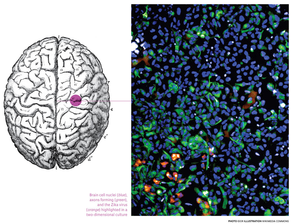
“We can investigate the action of the virus in increasingly complex cell models,” says neuroscientist Erick Loiola, postdoctoral research fellow at the D’Or Institute for Research and Education (IDOR) in Rio de Janeiro and one of the authors of the paper. He is referring to two-dimensional neural stem cell cultures grown in lab dishes; neurospheres, which are clusters of these cells; and brain organoids. The last of these, a kind of “mini-brain,” are more complex structures that mimic the features of a somewhat more-developed brain, like that of a fetus at three months. Loiola is part of a team led by neuroscientist Stevens Rehen, professor at the Federal University of Rio de Janeiro (UFRJ) and IDOR research director.
Since 2009, under the leadership of Rehen, researchers have been culturing lineages of induced pluripotent stem (iPS) cells that can produce a variety of tissues from adult cells, most often skin cells (see Pesquisa FAPESP Issue nº 156). Their work soon led to a technique for making the three-dimensional versions that serve as models in researching various facets of the functioning and development of the brain, like the psychiatric disorders that Rehen has been exploring for years.
Brain organoids have served as models in the study of microcephaly since 2013, two years before Zika won worldwide fame for its association with the birth of babies with below-normal head circumference for their gestational age and other neurological damage. Since IDOR was already producing these miniature brains, along with neurospheres, the lab was well positioned to respond to the epidemic and test the effects of the virus (see Pesquisa FAPESP Issue nº 242). Last year, the team found that neurospheres degrade when infected by the African strain of Zika and that infected cells have trouble forming neurospheres.
Altered DNA
The time had come to take a look from the perspective of the control room – that is, of genes – and conduct a more in-depth examination of what happens during the growth of cells infected with the strain of the virus circulating in Brazil. Cells isolated from a male patient in Espírito Santo were used to analyze cell RNA and its products (proteins), which are indicators of genetic activity. “We analyzed the neurospheres early on in their development, before the cells started dying off,” explains biologist Juliana Minardi, postdoctoral fellow at the Neuroproteomics Laboratory of the University of Campinas (Unicamp), which is headed by biologist Daniel Martins-de-Souza. They were already partnering with IDOR on a project about schizophrenia, so it was easy to apply the same model to Zika infection.
The idea was to investigate what was causing the observed impairment of neurosphere growth, when progenitor cells differentiate into the neurons and glial cells that form the brain structure. When researchers analyzed the difference between infected and healthy cells in the early days of the study, they detected changes in the expression of some 500 proteins, a range broad enough to trigger alterations in a gamut of cell functions. After identifying the proteins on the basis of earlier research, it became clear that Zika breeds an environment of instability that triggers the production of proteins linked to DNA repair. What then happens, however, is that these proteins inhibit the production of the molecules associated with the formation and differentiation of neural cells, while they also replicate the genetic material of the virus. The result is fewer, smaller, misshapen neurospheres. “The environment that is fostered by the virus in the neurospheres disrupts the normal life cycle of the cells, including neuronal differentiation,” concludes Martins-de-Souza.
For Unicamp researchers, this kind of close-up examination can help identify potential targets for drugs, both those currently undergoing testing for other diseases and others that have yet to be discovered. One example of a known medicine is chloroquine, used for decades to fight malaria. Tests with human neural cells and mice neurospheres showed that the drug can block viral proliferation and cell death, according to a study led by virologist Amilcar Tanuri of UFRJ, in partnership with Rehen’s team. Findings were published in the journal Viruses in December 2016 (see Pesquisa FAPESP Issue nº 245). Another collaborative effort, led by biologist Thiago Moreno Souza, of the Oswaldo Cruz Foundation (Fiocruz) in Rio de Janeiro, evaluated sofosbuvir, an antiviral drug normally used to treat hepatitis C. Results showed that it has a protective effect in organoids, as reported in Scientific Reports on January 18, 2017. Loiola, who also co-authored the paper, believes the results are highly promising, although not all countries have approved the use of sofosbuvir during pregnancy, when fetuses may become the greatest victims of microcephaly. “The lab is ready to offer the three neural models, which differ in complexity and speed of response, for use as a drug testing platform,” he says.
Project
Developing a predictive test for a successful medication response and understanding the molecular bases of schizophrenia through proteomics (nº 2013/08711-3); Grant Mechanism Young Investigators; Principal Investigator Daniel Martins-de-Souza (Unicamp); Investment R$ 4,774,285.05.
Scientific articles
GARCEZ, P. P. et al. Zika virus disrupts molecular fingerprinting of human neurospheres. Scientific Reports. V. 7, No. 40780. January 23, 2017.
SACRAMENTO, C. Q. et al. The clinically approved antiviral drug sofosbuvir inhibits Zika virus replication. Scientific Reports. V. 7, No. 40920. January 18, 2017.
DELVECCHIO, R. et al. Chloroquine, an endocytosis blocking agent, inhibits Zika virus infection in different cell models. Viruses. V. 8, No. 12, p. 322. December 2016.