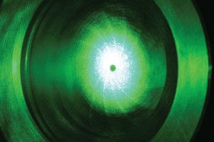
Miguel BoyayanLaser: information source about characteristics of biological tissuesMiguel Boyayan
The marriage of lasers with medicine is about to result in a tool that can improve the delicate work of screening livers to be used in transplants. Researchers from Cepof, the São Carlos Optics and Photonics Research Center of the University of São Paulo (USP) have developed a type of optic biopsy that informs, in real time and non-invasively, the amount of fat that has accumulated in the organ. The device that does the test – a very fine tube that emits a laser beam over the tissue under analysis, absorbs that light that is returned by the biological tissue and sends the data to an attached spectrometer and PC in a small case – was successfully tested on rats and, in a pilot study, on humans, by surgeons from USP’s Ribeirão Preto Medical School (FMRP).
The new approach informs, precisely and almost instantly, the degree of hepatic steatosis (lipids in the liver), an indispensible piece of information in the quest for the most viable organs for transplant. Fatty livers, with steatosis greater than 30%, cannot be used for this type of surgery. “This (the optic biopsy) may be a promising alternative to the conventional diagnosis methods used in surgeries and transplants,” wrote Giovanni Bottiroli and Anna Cleta Croce, from the University of Pavia (Italy) in an editorial in the journal Liver International, in March of this year. In the same edition of this international periodical, the scientist from São Paulo published an article reporting on their work with rodents. Cepof, where the liver fat diagnosis technique with lasers was developed, is one of the Cepids (Centers for Research, Innovation and Dissemination) financed by FAPESP.
The physician Orlando Castro and Silva Junior, head of the digestive surgery division and coordinator of the FMRP transplant group, is convinced that the new approach will be highly valuable in his area. “With the result of the biopsy in our hands, we just need to do a simple rule-of-three calculation to determine the liver’s degree of steatosis,” explains the surgeon, one of the authors of the work on animals and humans. There is rarely enough time to take a tissue sample from a liver available for transplant and to submit it to the requisite tests. Therefore, the diagnosis of the degree of liver steatosis is normally conducted subjectively by the surgeon him(her)self. “Today, assessment of the donated organ is conducted through the eye of the doctor,” comments Castro and Silva. “There is no time to perform a (conventional) biopsy.” Lasers may help shine a light into this gap of uncertainty.
To devise a fast and accurate diagnostic system regarding the accumulated fat in the liver, the USP researchers explored a very well-known physics phenomenon: fluorescent spectroscopy. Certain molecules can absorb energy and emit light after they are illuminated by a source of light. In the case of the work with hepatic steatosis, a laser beam applied to the biological tissue in question works as a fluorescence inducer. “What we did was to correlate the intensity of the fluorescence directly with the liver’s fat content,” explains Vanderlei Salvador Bagnato, Cepof’s coordinator and one of the main researchers in this field. “Each degree of steatosis (light, moderate or advanced) produces a different response.”
This strategy design is not new. The scientists from São Carlos had themselves worked along a similar line when they developed real time diagnostic systems using fluorescent spectroscopy for other clinical conditions, such as mouth and liver tumors, and the citrus canker that attacks orange plantations. In this type of approach, the first step is to find out whether the technique captures the biological changes being studied and if it can generate a reliable standard that allows one to differentiate the different stages or phases of this pathology. “We have to find out if the method is sensitive to this biological condition in an animal model,” says Cristina Kurachi, one of the Cepof researchers. “Without this confirmation, it is very difficult to propose the technique for use on humans.”
The tests on rodents encouraged the researchers from São Carlos and Ribeirão Preto – as well as the editors of the journal Liver International. The fluorescence spectroscopy diagnosis also managed to separate the rats into the four large groups that they belonged to in clinical terms: normal animals (with no liver fat), and animals with low, moderate and high hepatic steatosis. In the article in the medical journal, the USP team shows that, by directing a laser beam of a given wave length onto the biological tissue, a steatosis fluorescence factor is generated that varies linearly with the amount of liver fat: the more the fat, the greater the factor. With this information, surgeons who are unsure about the real condition of a donated liver can promptly discover the organ’s fat level. “We tested the method on 27 donated human livers, six of which have already been described in our pilot study, and it seems to be fantastic for diagnosing steatosis,” states surgeon Castro and Silva. “I believe that we can use this technology to analyze other liver problems, such as cirrhosis.” Before being used in operating rooms, however, lasers in the service of medicine will have to be proven in more human livers destined for transplants.
The project
Biophotonics – optical fluorescence; Type Cepids (Centers for Research, Innovation and Dissemination); Coordinators Vanderlei Salvador Bagnato and Cristina Kurachi – Cepof (São Carlos Optics and Photonics Research Center at the University of São Paulo – USP) Orlando Castro and Silva Júnior – Ribeirão Preto Medical School from the University of São Paulo; Investment R$ 40,000 and US$ 32,000 a year for the project (FAPESP)
Scientific article
Oliveira, G.R. et al. Fluorescence spectroscopy to diagnose hepatic steatosis in a rat model of fatty liver. Liver International. v. 29, n. 3, p. 331-336. Mar. 2009.