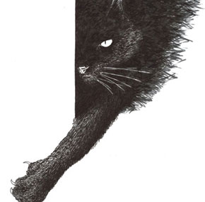
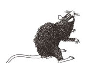
 DANILO ZAMBONISet a rat in front of a cat and you will witness one of the reactions that are key to survival. The rat will promptly become immobile, as if it were dead, thereby diminishing the risk of catching the eye of its predator, generally attracted by movement. If the danger persists, however, or if the cat moves closer, the rodent starts jumping backward vigorously, which enables it to fly through a distance equal to several times the length of its body. The freezing of the movement and the energetic attempt to flee are part of the repertoire of natural defense reactions that are typical of situations that trigger fear. They probably arose hundreds of millions of years ago, with the first reptiles, which escaped from their predators and spread over the Earth and they continue to be found among a broad group of animals that includes mammals – human beings amongst them. However, it was only recently, based on studies conducted in Brazil and in the United States, that scientists found that the reactions that prepare the body to fight or flee in the face of danger are triggered and coordinated by a deep and primitive area of the brain: the hypothalamus, a structure with the shape and size of an olive, located at the base of the brain, at the height of the eyes.
 DANILO ZAMBONI
Intrigued by the number and the complexity of the changes that defense reactions trigger in the body, momentarily rising the blood pressure, heightening attention and preparing the muscles to spring into action – the physician and neuroanatomist Newton Sabino Canteras decided, slightly more than a decade ago, to venture into the complex neural circuits of the hypothalamus. Protected in humans by the brain’s hemispheres, this structure slightly more than 2 cm long, 1 cm wide and almost 2 cm high has at least 16 different sets of cells, with connections among themselves as well as with other parts of the brain and with other organs in the central nervous system. It produces various hormones and is linked to the control of hunger, thirst, body temperature, sleep, reproductive behavior and aggressiveness.
The careful investigation into how these circuits connect inside and outside the hypothalamus and the determination of the sequence in which they are activated in situations that put life at risk, such as a predator’s attack, led Canteras and researchers from the United States to propose that this brain structure plays a key role in the generation and the coordination of the defense reactions triggered by fear and in the recollection of the circumstances that gave rise to it. Experiments conducted at Canteras’ laboratory at ICB, the Biomedical Sciences Institute of the University of São Paulo (USP), also made it possible to see that unlike what was previously believed, the path of fear in the brain is not a single one: fears triggered by different situations my activate different cell circuits.
More than a detail of rat brain physiology, the discovery that certain sets of cells in centers of the hypothalamus are activated in reaction to fear may even influence our understanding and treatment of mental conditions such as anxiety, which afflicts 4% of all Brazilians, and its more extreme and uncommon version, panic, which leads 1.6% of the population to suffer sudden crises of shortage of breath and tachycardia, during which the mind is taken over by the certainty that one is about to die. There is good reason to review current knowledge of such problems: a lot of what is known about how these conditions arise and evolve is based on experiments with animals (rodents in particular), simulating threatening situations that are different from those encountered in nature.
In research laboratories, the scientists try to reproduce risk of death situations by giving a rat light electric shocks on its paw, under certain circumstances. This type of threat, however, seems to lack the same evolutionary meaning as predators. Over the course of thousands or even millions of years, the animals did not have to deal with electrical discharges such as those in the forests, savannas and deserts. The shocks may frighten and bother them, but they cause no lesions. Generally, in these environments, the animals faced a rather different reality: while looking for food, they had to flee from larger or stronger animals that were bent on turning them into a meal. This is more or less what happens with a rat that, upon leaving its den, runs straight into a cat, or what happened to the forebears of Homo sapiens when they left their cave in search of food. “The experimental model that resorts to shocks to simulate the effect of fear is very artificial,” states Canteras. “The shock gives rise to an aversion, but it doesn’t awaken fear as do the situations that put survival at risk.”
To understand how the response to fear is processed in the brain, Canteras chose, some 15 years ago, to use a truer representation of what supposedly occurs in nature. With the couple Robert and Caroline Blanchard, from the University of Hawaii, in the United States, he developed an experimental model in which a rat spends a few minutes in front of a cat. Even though it is protected within a transparent acrylic box, safe from the feline’s claws, the rodent usually starts to flee as if there were nothing between it and its predator.
Right from his early experiments, Canteras found that merely exposing the rat to the cat increased the level of activity in certain parts of the rat’s hypothalamus. More specifically, it activated a group of cells called the dorsal pre-mammillary nucleus. Although it contains only a small set of cells – just a few thousand neurons out of the billions that are found in the brain – the dorsal pre-mammillary turned out to be essential for defense reactions. Without these neurons, instead of lying still or trying to spring away, the rats would leave the acrylic box to explore the environment despite the cat, as if the predator were not there. They would often approach the cat as if they no longer feared it, Canteras showed in 1997 in an article published in the Brain Research Bulletin, which he wrote together with Silvana Chiavegatto and Luiz Ribeiro do Valle, both from ICB, and Larry Swanson, from the University of Southern California, in the United States.
It has been known since the 1920’s that the hypothalamus is in some way involved in defense behavior. Tests conducted at that time by the US physiologist Archibald Bard with cats suffering from brain lesions indentified an area at the back of the hypothalamus that was important for the manifestation of the so-called fictitious anger – a defense hyper-reaction associated with the severance of the connections between the deep areas of the brain and the cortex, which is the most superficial one. In subsequent decades, other centers of the hypothalamus were mapped, but nothing was known about the function of the dorsal pre-mammillary nucleus until the 1990’s. During the time he spent at Swanson’s laboratory from 1990 to 1992 Canteras dissected the connections of the nuclei of the medial (central) part of the hypothalamus – including the dorsal pre-mammillary one – connected with the expression of innate or learnt behaviors. After returning to São Paulo, he initiated tests to understand how these areas of the hypothalamus worked.
At Canteras’ laboratory, the psychologist Alessandro Cezario and the biologist Erika Ribeiro Barbosa, along with Marcus Vinicius Baldo,from the ICB Sensory Physiology Laboratory, conducted a more elaborate series of tests. They placed the rat inside an acrylic box and food inside another box, connected to the first one by a tunnel. In an initial stage, the rat had to go through the tunnel to reach the food. However, upon arriving at the place where the ration was meant to be, the rat would find a cat. As a result, it would become paralyzed. This was the expected defense reaction to the innate or non-conditioned fear of the rodent vis-à-vis its predator – something akin to what presumably occurs with someone who upon going round a corner walks straight into a fierce-looking dog.
In the second series of tests, the rodent had to cover the same path to eat,but the cat was no longer in the food compartment. Even so, upon entering the environment where it had seen the feline, the rat froze for a while, until it eventually started cautiously exploring the place – like someone who, having been bitten by a dog in front of a given house, starts avoiding that sidewalk for a while.
The aim of the experiment was to see what happened with the hypothalamus in situations that cause conditioned fear, i.e. when one can anticipate danger. In this case, the dorsal pre-mammillary nucleus also turned out to be the most active region. If, however, this group of cells was destroyed before the first phase of the experiment (when the rat runs into the cat), the rodent lost its fear of exploring the place on the following day, when the feline was no longer there, as shown by the researchers in a article from 2008 in the European Journal of Neuroscience. This was a sign that the rodent had lost the capacity to recall the terrifying meeting and to associate it with the environment in which it had taken place. In an article published online in January of this year in Neurobiology of Learning and Memory, the Canteras team showed that the information travels from the dorsal pre-mammillary nucleus to a center that stores memories associated with emotions, such as fear.
By injecting directly into the dorsal pre-mammillary nucleus compounds that block the working of the neurons the ICB group found that both in the first and in the second case the interruption of the activity of this region reduces – and by a great deal – the activation of a neighboring area: the periaqueductal gray matter. Located in the mesencephalon (a structure that lies between the brain and the spinal cord), the periaqueductal gray matter controls the cardiovascular and behavior changes that the animal has in the face of its predator. It also leads to the release of analgesic substances and others that raise the anxiety level. An experiment described in 2007 in Science mapped the activity of the periaqueductal gray matter among people who were playing a virtual game in which they were pursued by a predator and showed that the functioning of this brain region intensified as the threat came closer and despair rose. Corroborating these observations, the recent work of Cristina Del-Ben and Frederico Graeff, from USP in Ribeirão Preto, suggests that alterations in this part of the mesencephalon may form the neurobiological basis of panic syndrome.
It is now clear that the sound functioning of the dorsal pre-mammillary nucleus is fundamental for the generation of defense reactions in the face of danger (whether real or potential). Without it, one loses the ability to feel fear, which is as instinctive and essential for the survival of any individual and its species as the need to eat and procreate. “This nucleus works like an amplifier of signals related to the presence of a predator,” explains Canteras, who, along with Antonio Carobrez, from the Federal University of Santa Catarina, found that noradrenalin is an important chemical communicator released in this region in situations involving fear.
Prior to these studies, tests conducted by the psychologist Joseph LeDoux, from New York University, attributed the coordination of responses to fear to the amygdala, an almond-like structure that first appeared among early mammals, in the temporal lobe. It is a known fact that the amygdalae (there is one in each side of the brain) receive visual, audio and smell information about the threatening situation. Previously, scientists believed that they directly activated the neurons of the periaqueductal gray matter, responsible for the changes that prepare the body to fight or flee.
Canteras’ work, however, indicates this is not quite the way this works. The stimuli that the predator awakens and those that are linked to the environment in which the prey and predator found themselves are, at first, compiled by the amygdala. However, they then converge toward the hypothalamus, where they are processed in the dorsal pre-mammillary nucleus, which relays them to the periaqueductal gray matter.
These observations, once combined, indicate that it could be necessary to review and refine the experimental models that have been used for decades to understand how the brain reacts to fear. The models are based on ideas put forth in the 1970’s by the American psychologist Robert Bolles, according to whom the fear caused by real threats (predator) and the fear caused by artificial situations (shock) activate the same parts of the brain in so far as they generate the same type of response.
Recently, Canteras obtained yet one more indication that the path of fear in the brain may vary depending on the type of threat. Canteras and Simone Motta,from ICB, along with Swanson planned an experiment in which the aggression did not come from a predator, but from another rat. For a few minutes, they would put a rodent in the cage of a stronger male that lived with a female. Right at the start the stronger rat attacked the intruder. The latter, in response, became paralyzed, which signals submission. This danger situation also involves activating the hypothalamus and the periaqueductal gray matter, but from regions that are not the same as those that were activated in the face of the predator, stated the researchers in a 2009 article in the Proceedings of the National Academy of Sciences. “In this case,” Canteras tells us, “the activity of the circuit in the hypothalamus that is related to social interactions rises.”
The project
Neural bases of motivated behavior Bases (nº 05/59286-4); Type Thematic project; Coordinator Newton Sabino Canteras – ICB/USP; Investment R$ 1.173.284,17
Scientific articles
MOTTA, S.C. et al. Dissecting the brain’s fear system reveals the hypothalamus is critical for responding to subordinate conspecific intruders. PNAS. v. 106, n. 12, p. 4,870-875. 24 March 2009.
CEZARIO, A. T. et al. Hypothalamic sites responding to predator threats – the role of the dorsal premammillary nucleus in unconditioned and conditioned antipredatory defensive behavior. European Journal of Neuroscience. v. 28, n. 5, p.1.003-15. 2008.