
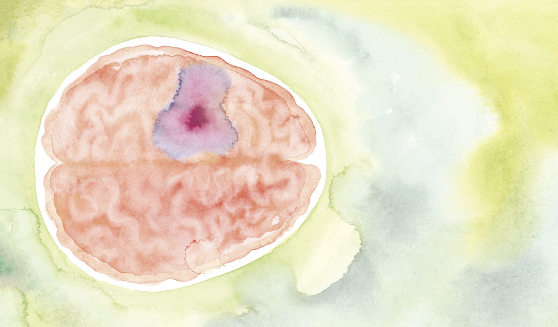
How is it that tumorous cells already grown in organs such as the lungs, kidneys, and intestines are able to get to the brain if it is protected by barriers that should have halted their advance? Researchers from the states of Alagoas and São Paulo have found a possible explanation: tumor cells emit chemical signals that act on the central nervous system cells and allow them to pass through the walls of the blood vessels and install themselves in the encephalon, a structure with 86 billion neurons formed of the cerebellum, at the base of the brain, and of the brain itself. Once in the brain, these invaders are protected from the defense cells, which, in other parts of the body, would attempt to destroy them.
This mechanism emerged through observation of molecular interactions between three types of cells: tumor, astrocyte (brain cells that nourish and sustain the neurons), and the blood vessel cells of the blood-brain barrier, a type of membrane normally preventing entry into the brain of molecules, cells, and impurities capable of adversely affecting neuron function. The conclusions are supported by analyses conducted using a technique known as single-cell RNA sequencing, which reveals the molecular behavior of each cell. Examinations were conducted on 128,421 cells extracted from 36 samples of brain tumors arising from the metastasis (spread) of cancers originally initiating in the breast, lungs, skin (melanoma), kidneys, ovaries, and intestine.
Brain metastases represent 6% of all cancer cases per year, and are 10 times more common than tumors originating in the brain itself. It is estimated that in 20% of people with cancer—primarily of the lungs, breast, intestines, and kidneys—the tumor cells will move from the organs in which they were formed and arrive at the brain. In these cases, even with the various possibilities for medical intervention, the survival rate after diagnosis is low.
“Using a technique known as spatial transcriptomics, which maps areas where specific genes are active in the brain tissue, we were able to see exactly where each cell interacts with the surrounding environment,” says biologist Carlos Fraga, of the Federal University of Alagoas (UFAL), Arapiraca campus, lead author of the article presenting the results, published in March in the journal Brain Research. Coauthor of the study, immunologist Helder Nakaya, of the Albert Einstein Israelita Institute for Education and Research and the University of São Paulo’s School of Pharmaceutical Sciences (FCF-USP), adds that the analysis methods also indicated which cells are close to one another, and how they arrange themselves in the tumoral environment.
According to this study, tumor cells produce a protein: vascular endothelial growth factor (VEGF), which acts on the cells of the blood-brain barrier vessels and stimulates the formation of new vessels wherever they advance. Other compounds increase the vascular permeability, facilitating the passage of the tumor cells, which then release other substances, including integrins, that arrive at the astrocytes and enable the invaders to go unrecognized as malignant, install themselves among the nervous cells, and resume multiplication. Specific genes, such as ITGA8, NRP1, and PLXND1, act as mediators of the interaction between tumor cells and the blood-brain barrier.
“An important contribution of this study was the identification of common mechanisms used by tumor cells of different origins to pass through the cerebral barrier,” comments physician Roger Chammas, director of the Center for Translational Oncology Investigation at the São Paulo State Cancer Institute (ICESP), who did not participate in the study. “These discoveries still need to be validated by wider studies, but already represent a significant advance.”
The team responsible for these findings intends to analyze the so-called primary tumors, formed in the brain itself, such as glioblastomas, as soon as possible. “We want to compare primary tumors with brain metastases to better understand the route taken by tumor cells, from the tissue in which they are formed, passing through the bloodstream, and installing themselves in the brain,” explains Nakaya.
The story above was published with the title “The brain invasion” in issue 351 of May/2025.
Projects
1. Systems biology applied to transcriptomic analysis of inflammatory diseases (n° 22/02605-6); Grant Mechanism Postdoctoral Fellowship; Supervisor Helder Takashi Imoto Nakaya (USP); Beneficiary Carlos Alberto de Carvalho Fraga; Investment R$148,840.95.
2. Integrative biology applied to human health (n° 18/14933-2); Grant Mechanism Young Investigator – Phase 2; Principal Investigator Helder Takashi Imoto Nakaya (Albert Einstein Israelite Institute of Education & Research); Investment R$2,309,586.43.
Scientific articles
DE CARVALHO FRAGA, C. A. et al. Revealing shared molecular drivers of brain metastases from distinct primary tumors. Brain Research. Vol. 1851, 149456. Mar. 15, 2025.