 Mariana CoanStarting to get a better understanding of the reason that people with Down syndrome, which affects one in every 700 children, are more susceptible to develop immune-system diseases than the rest of the population. In a study published in September in the Journal of Immunology Brazilian researchers showed that in the case of these children, a sophisticated mechanism that teaches the defense cells to recognize and combat what is foreign to the organism, is out of synch.
The result of this imbalance is that the cells which should protect the body begin instead to attack it, causing it to develop immune-system diseases such as Type 1 diabetes, hypothyroidism or celiac disease.
The pediatrician Magda Carneiro-Sampaio and her team at the Instituto da Criança (Institute of the Child) (ICr) of the University of São Paulo’s School of Medicine (FMUSP) verified that something was not right with the maturation of the defense cells of children with Down syndrome when they compared the activity levels of their thymus glands to those of children who did not have this problem.
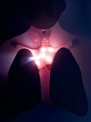
A small flattened organ in the shape of a butterfly, the thymus gland is located in the thorax, behind the sternum and in front of the heart, and operates like a school for war training. It’s here that a special group of defense cells – the T lymphocytes, which are responsible for orchestrating the fight against infections and the elimination of diseased cells – learn to distinguish between what is a part of the body – and should be preserved – and that which is foreign to the organism – and should be exterminated.
When the thymus gland is functioning properly, the lymphocytes that undergo this training and show themselves capable of recognizing and attacking the cells of the organism itself, they are destroyed there and then – this is the fate of between 95% and 97% of the T lymphocytes. Only the remaining 35 to 5% of the lymphocytes, demonstrating that they have the ability to identify and attack only infectious agents and compounds that are foreign to the body and defective cells, are allowed to leave the thymus gland and make their way into the bloodstream and the lymphatic system. However, in the case of people suffering with Down syndrome, this rigorous cell preparation and selection system is out of synch.
The imbalance in the maturation of the lymphocytes has only begun to become clear in the last few years, when Magda’s group used molecular biology techniques to study the thymus glands of 60 children (14 with Down syndrome and 46 without) aged between 4 months and 12 years. All of them had undergone surgery to correct serious heart defects that demanded that the thymus gland be removed. By making a comparison of the functioning of the thymus glands the researchers were able to show that, on average, this organ was less active in the case children with Down syndrome than it was in those who did not have this problem (see info graph above).
The geneticist Carlos Alberto Moreira Filho and the psychiatrist and specialist in bioinformatics Helena Brentani assessed the level of activation of almost 22 thousand genes in the cells of the thymus gland and verified that 400 of these genes, many of them responsible for cellular multiplication and for the maturation of the defense cells, were less active in children with Down syndrome.

One in particular caught their attention. This is the autoimmune regulator gene (AIRE). This gene encodes the production of a protein that is essential for the proper selection of the T lymphocytes. Without this protein, the lymphocytes that are harmful to the organism itself are not exterminated inside the thymus gland, as they should be, and instead spread throughout the body.
The pathologist Maria Irma Seixas Duarte and the biomedical researcher Flavia Afonso Lima observed that there were twice as many cells with the AIRE gene that were active in the thymus gland of children without Down syndrome as there were in the case of children with this problem. On average, 155 cells per square millimeter were releasing the AIRE gene in the thymus glands of children in the first group, while the comparable figure was one of 70 cells in the case of the second group. “The low registration level of the AIRE gene makes it possible to understand why auto-immune diseases are more common among those with Down syndrome,” explains Magda.
The pattern in terms of the activation of the genes in the cells of the thymus gland also help to explain the clinical signals observed in children with Down syndrome, the most common chromosome anomaly in human beings, which is caused by the presence of an extra copy of chromosome 21 in the core of the cells. From very early on in life, a large percentage of those people who have Down syndrome present auto-immune problems that have been triggered by defense cells attacking specific organs.
The risks of developing hyperthyroidism, type-1 diabetes or celiac disease are 4 times, 6 times and between 10 and 40 times greater, respectively, among children with Down syndrome, than they are among the rest of the population. For almost three decades now, it has also been known that the thymus glands of children who have this syndrome are smaller than those of children without the chromosome anomaly.
In light of these new results, Magda and her team propose to carry out a reinterpretation of the origin of the auto-immune problems that are common among those who have Down’s syndrome. “The infirmities that these children present are the result of a primary immunodeficiency, rather than the current classification, which is that of secondary one,” she states.
What does this reevaluation mean? First, that the cause of the auto-immune diseases in the case of people with Down is different to what was previously thought. “The origin of the poor functioning of their defense system is genetic and appears during the formation of the embryo,” explains Magda. Up to now, the explanation that was most-widely accepted by the specialists was that these problems in the auto-immune system were the result of the degeneration of the thymus gland due to premature ageing. And the second most accepted explanation was these children might not be receiving the proper medication.
 Mariana CoanIn order to improve the treatment of these children, in December at the Darcy Vargas Children’s Hospital, in São Paulo, the team of Magda and of the pediatrician Zan Mustacchi initiated the triage of those who have Down syndrome and who exhibit recurring infections even after having been vaccinated against viral and bacterial diseases.
They intend to verify whether this increased susceptibility to infections – they can aggravate the heart problems that are commonly observed in children with Down syndrome – are also the due to the poor functioning of the thymus gland. “If this is confirmed, we will be able to program a supplementary vaccination in an attempt to improve the immune response of these children and, in certain cases, to indicate the preventive use of anti-virals and antibiotics for those have congenital cardiopathy,” says Zan.
Years ago the group from the Instituto da Criança decided to investigate the activity of the thymus gland in Down syndrome because the pattern of immunological problems exhibited by these children recalled another rare infirmity that is associated with the improper functioning of this organ: type 1 auto-immune polyendocrinopathy (APECED). Common among Italians from Sardinia, Finnish people and Iranian Jews, this polyendocrinopathy, is also characterized by the abnormal activity of the thymus gland. In both cases, lymphocytes that should be destroyed escape selection and attack the body itself due to the abnormal activity of the AIRE gene, which is found in chromosome 21.
In the case of APECED alterations in the structure of this gene, such as were found by Magda’s group in 2007 in a Brazilian family of Italian origin, have a negative impact on the production of the AIRE gene and the selection of the T lymphocytes. In the case of Down syndrome, very small sections of genetic material – the micro-RNAs, which are found in abundance in chromosome 21 – can interfere in the activity of AIRE and of other genes. “We intend to investigate the role that these micro-RNAs play in the next stage of the research,” declares Magda.
She and the USP researchers are also planning to use tests that make it possible to evaluate the thymus gland’s size and activity level in order to identify, if possible even before the birth, these and other severe primary immunodeficiency. Considered to be rare, these infirmities show up very early in life and make the children more susceptible to infections and auto-immune problems. It is calculated that one out of every 10 thousand children presents some type of severe immunodeficiency (part of the cases with alteration in the thymus gland), that are almost always fatal without the proper treatment.
One of the immunodeficiencies that the researchers hope to detect early is that of DiGeorge syndrome, which affects one out of every 4 thousand children. The result of the loss of a piece of chromosome 22, this syndrome causes defects in the heart and in the face and prevents the normal development of the thymus gland. Between 1% and 2% of the children with this syndrome may even be borne without the thymus gland, which impedes the formation of the immune system and can only be corrected by means of the transplant of this organ. The ICr also wants to identify the cases of severe combined immunodeficiency, which affects 1 out of every 40 thousand babies born.
In recent years, a number of states in the US have included the Guthrie Test in the neonatal screening. This test is an exam that measures the number of lymphocytes that were recently released by the thymus gland, which functions as an indicator of the organ’s activity in the blood. However, this genetic test is still too expensive to be adopted by the public health system of countries such as Brazil – it would cost US$ 2.4 million a year to apply the test to the 600 thousand children who are born each year in the State of São Paulo.
For this reason, the USP group is thinking of taking advantage of the ultrasonography of the fetus, which is done during gestation, in order to assess the size of the thymus gland. “It would just be one more item to be verified during the evaluation of anomalies that is carried out by ultrasound,” says Luiz Antonio Nunes de Oliveira, who is head of the ICr’s Radiology Service.
Since the thymus gland is relatively large in the fetus, it is possible to identify it by means of this imaging exam. “Those cases where the thymus gland is smaller, or is not visible would be regarded as suspect and the doctors could request a leucogram soon after birth,” explains Oliveira. The obstetrician Roseli Nomura, of FMUSP’s Department of Obstetrics and Gynecology, is now working on discovering the best technical conditions to assess the thymus gland by ultrasound at the final pre-natal exam, without significantly increasing the duration or the price of the exam.
Early identification of abnormal activity of the thymus gland is important for the survival of the new-born baby. For example, children with serious immune system deficiencies should not receive the BCG vaccine, which is applied shortly after birth. This anti-tuberculosis vaccine is produced with live bacillus, which can cause a serious infection – and even be fatal – for these babies.
“The earlier the diagnosis is done, the earlier one can program the proper immunization for the child,” affirms the pediatrician Cristina Jacob, head of ICr’s Allergy and Immunology. In the cases of serious combined immune deficiency, the early diagnosis enables the child to be forwarded rapidly for the transplant of haematopoeitic cells, which is the only therapy possible at the present time.
The Project
Autoimmunity in children: investigation of the molecular and cellular bases of the autoimmunity of early onset (nº 2008/58238-4); Modality Thematic Project; Coordinator Magda Carneiro-Sampaio – FMUSP; Investment R$ 1,470,770.68 (FAPESP)
Scientific article
LIMA, F. A. et al. Decreased AIRE expression and global thymic hypofunction in Down Syndrome. The Journal of Immunology. v. 187 (6), p. 3422-30. 15 Sep. 2011.