Abundant viruses contribute to the genetic diversity of life on Earth and may have emerged four times throughout the planet's history
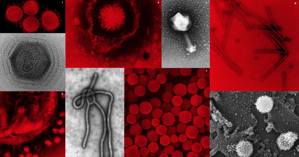
The many forms of viruses: bacteriophage (1); ebola virus (2); bombyx mori nucleopolyhedrovirus (3); tupanvirus (4); mimivirus (5); SARS-CoV-2 (6); herpes virus (7); myovirus (8); piryvirus (9); tobacco mosaic virus (10); influenza virus (11)
1 Graham Beards / Wikimedia Commons 2 Cynthia Goldsmith / CDC 3 Bergmann Ribeiro / UNB 4 Jônatas Abrahão / UFMG 5 XIAO, C. et al. PLOS Biology. 2009 6 NIAID / NIH 7 E. L. Palmer / CDC 8 SULLIVAN, M. B. et al. PLOS Biology. 2005 9 Fred Murphy / CDC 10 Wikipedia 11 Cynthia Goldsmith / CDC
For the last six months, there has been only one topic of conversation. Since it was identified in China in December 2019, the novel coronavirus has become ubiquitous. It has spread to all of the world’s major cities, infecting at least 6.5 million people and killing more than 380,000 in a pandemic that has moved at a frightening pace. Hospitals were quickly overwhelmed, leading to significant changes to our daily lives. It did not take long to reach even the most remote areas of the planet, from indigenous groups in the Amazon rainforest to the high-altitude, cold lands of Bhutan, in the Himalayas. The novel coronavirus, however, is just one of hundreds of thousands—perhaps millions—of virus species believed to exist, which, thanks to modern transport systems, are able to spread at speeds that would have been unimaginable a few decades ago. Faced with this situation, it is important to understand more about what viruses really are and how they emerged and spread to all corners of the earth. These tiny living organisms, composed of genetic material and proteins, can be capable of causing devastating diseases.
Recent research has helped with this task. In an article published in the journal Microbiology and Molecular Biology Reviews in March, an international group of virologists created the first comprehensive system for classifying viruses, and by grouping species according to their degree of genetic similarity, concluded that they have appeared at least four times in the planet’s 4.6-billion-year history. They initially arose long before the first cellular organisms appeared on Earth, at least 3.5 billion years ago. On the other three occasions, it is likely that rudimentary cell types already existed.
Defining the relationship between viruses by their degree of genetic proximity may seem like an obvious strategy, but there are two reasons why it was almost impossible until recently. Firstly, the number of viruses that have been studied and described is relatively small. The International Committee on Taxonomy of Viruses (ICTV), the body responsible for naming and classifying viruses, has only cataloged 6,590 species. This is almost nothing compared to the approximately 1.2 million species of cellular organisms known to science—a group that includes bacteria, archaea, protozoa, plants, and animals. Secondly, viruses have a small number of genes, very few of which are common among different species, which makes it difficult to compare and establish relationships between them.
The situation has begun to change in the last 15 years with a rise in metagenomics, a method through which scientists analyze genetic material recovered from environmental samples without the need to isolate and cultivate viruses in a laboratory. With this strategy, experts have already identified hundreds of thousands of new virus species, which are waiting to be described. “Metagenomics research has led to the discovery of species that have filled many of the gaps in the diversity of viruses—the virosphere—and has increased the reliability of evolutionary studies,” says Brazilian virologist Francisco Murilo Zerbini, from the Federal University of Viçosa (UFV) in the state of Minas Gerais.
Zerbini is a member of the ICTV’s executive committee and is coauthor of the new taxonomy, created under the coordination of one of the most respected scholars in viral evolution, Russian biologist Eugene Koonin, from the National Center for Biotechnological Information (NCBI) in the USA. The group reorganized viruses based on two criteria: the type of compound used to store genetic information—ribonucleic acid (RNA) or deoxyribonucleic acid (DNA)—and the level of similarity between certain genes, common among as many viruses as possible. The result is a comprehensive, cohesive, and robust framework for classifying viruses, the first since the existence of these infectious agents was first proposed in 1898 by Dutch botanist Martinus Beijerinck (1851–1931).
The new classification separates viruses into four large groups, called domains, which are added to the two others used to categorize all cellular beings. The domain, also known as a realm, is the highest of the taxonomic categories, above kingdom, phylum, class, order, family, genus, and species, which together group living beings in increasing order of similarity. A domain includes the largest possible number of species with just a few common features. To give an idea of their vastness, all living beings formed of cells (bacteria, archaea, protozoa, fungi, plants, and animals) are divided into just two domains: the eukaryotes, which encompasses cellular organisms whose genetic material is stored in a compartment called a nucleus, and prokaryotes, the cells of which have no nucleus. Since viruses have such a high degree of diversity, they had to be separated into four domains, but this is still an improvement over previous classification attempts, which have tried to group them according to anatomy, the type of tissue to which they are chemically attracted, or the type of genetic material.
In the new system, viruses are grouped into the domains Riboviria, Monodnaviria, Varidnaviria, and Duplodnaviria. Riboviria includes all viruses that store information about their structure and how they function—their genes—in an RNA molecule. “Many researchers believe that life began in an aquatic environment where RNA molecules stored genetic information, known as the RNA world,” explains Zerbini. “The theory is that RNA viruses descended from that world, appearing before cellular organisms.”
This group includes hepatitis, colds, influenza, dengue, HIV, and the novel coronavirus SARS-CoV-2. Composed of the nitrogenous bases adenine (A), cytosine (C), guanine (G), and uracil (U), RNA is usually found in nature in the form of a single strand, although viruses can also be double-stranded. It is more malleable and can perform more biological functions than its close relative, DNA, which has a thymine (T) base instead of uracil and can have a single or double strand. DNA acts as a kind of instruction manual on how to produce a new virus, cell, or multicellular organism. RNA can play this role too, but it can also function as an enzyme, accelerating chemical reactions, or as a messenger, translating instructions on how to make proteins and delivering them to a cell’s protein factories.
The viruses in the other three domains store their genetic information in DNA: on a single strand in the case of Monodnaviria, or a double-strand, like cellular organisms, as occurs with Varidnaviria and Duplodnaviria—the difference between which is a capsid protein, the shell that protects genetic material.
Identifying these four domains allowed the researchers to draw conclusions about the evolutionary history of viruses. Each domain is composed of species that descend from a common ancestor that existed billions of years ago. Four domains means four ancestors, which must have arisen at different times, given their distinct genetic characteristics. “The data available so far indicate that based on their evolutionary relationships, it is not possible to unify all viruses in one category or to infer that there was a common ancestor to all viruses,” says Zerbini.
Having arisen numerous times over history is another feature that distinguishes viruses from cellular organisms. Analyses of some 350 genes common to cellular organisms (from tuberculosis bacteria to ferns; from amoebas to mountain gorillas) indicate that they all descend from a common ancestor, a single-celled being that existed between 3.5 billion and 4.5 billions of years ago.
Viruses are almost always placed in a separate group to cellular organisms. Some virologists say that as a result, they are usually defined by a process of elimination: they are not bacteria, they are not fungi, they are not plants, and not animals. But this was not always the case.
Martinus Beijerinck, the Dutch botanist who first used the term virus to name an infectious agent, proposed that they were living beings. He repeated experiments carried out in 1892 by Russian botanist Dmitri Ivanovsky (1864–1920), who filtered diseased tobacco leaves through a material with very fine pores capable of retaining the smallest known forms of life at the time (fungi and bacteria). Like the Russian, Beijerinck found that the remaining liquid was capable of causing disease, and that the pathogen it contained reproduced in proliferating cells. In an 1898 presentation to the Amsterdam Academy of Sciences, he called the liquid a contagious living fluid or virus (poison or toxin, in Latin) and stated that it contained an infectious agent of an unknown nature.
Later studies of these agents showed that they were composed of proteins and a small number of nucleic acids—RNA or DNA, the components of genes. Proteins, nucleic acids, sugars, and fats are the four groups of chemical components found in all cellular organisms. Although viruses have two of them, they were originally considered merely as protein structures, which seems to have contributed to the notion that they were not alive.
One famous example of viruses being excluded from the living world is the tree of life proposed in the 1970s by American biologist Carl Woese (1928–2012), which only includes beings formed by cells.
Experts like Koonin, who consider viruses to be alive, believe that part of the prejudice stems from the fact that for a long time, they have only been studied outside the cells, when they are inert. An indisputable fact among both virologists and biologists, the latter of whom generally study cellular organisms, is that such beings were the first forms of autonomous life to appear on Earth.
Cells are like microscopic pouches containing encrusted proteins, with a double outer layer made of a type of liquid fat (lipids). Only cells are equipped with the components needed to independently perform the three phenomena almost always associated with life: generating energy, reproducing, and accumulating changes from one generation to the next—evolving. Viruses, regardless of when and how many times they have emerged on Earth, actually accumulate genetic changes much faster than other organisms, but they can only obtain energy and replicate themselves if they are inside a cell, which is almost always enslaved to the virus. Outside cells, they are inert and harmless.
“Viruses exist in a gray area between the living and nonliving, at the line where chemicals become life,” says virologist Eurico Arruda, from the University of São Paulo (USP) in Ribeirão Preto. Arrua, who specializes in viruses that cause respiratory diseases, recently found that the influenza virus does not only attach to the cells that line the nose, mouth, throat, and respiratory tract. According to an article published this year in the Journal of Virology, it also infects immune system cells in the tonsils and adenoids, located in the throat. Influenza A can lie dormant in these cells for long periods, but still be able to cause infection. “Viruses are very elegant biochemical parasites, capable of infiltrating a cell’s command center and using the cellular apparatus to propagate and evolve, causing biological side effects. For me, this is life,” he says.
Many virologists find the issue either inconvenient or unimportant. French biologist André Lwoff (1902–1994), one of the founders of the ICTV and winner of the 1965 Nobel Prize in Physiology or Medicine for explaining how viruses insert their genetic material into the DNA of bacteria, has said in interviews that the question does not matter and that whether they are alive or dead is a matter of opinion. Others are more pragmatic. They say that inside cells, viruses are alive. Outside, they are not. There are also some, such as French biologist Patrick Forterre of the Pasteur Institute in Paris, for whom the problem lies in the definition of life and organisms, which should be expanded to better describe that found in nature.
One of the arguments is the abundance of viruses, which are found in all environments. Genomic studies suggest that the number of viruses on the planet far exceeds that of all other living beings put together. According to seawater sample analyses, there are 3 million to 100 million virus specimens in just one milliliter of seawater. Extrapolating that figure worldwide, the oceans contain an unimaginable total of 1031 viruses, 10 billion times more than the total number of stars in the universe. In an article published in the journal Nature in 2005, virologist Curtis Suttle, from the University of British Columbia, Canada, calculated that if they were all laid end to end, these viruses would stretch almost 100 times further than the diameter of the Milky Way, despite being submicroscopic—they are 30 to 600 nanometers in diameter. “Because they infect and often kill microscopic algae, viruses can reduce oxygen production in the oceans by 3% to 4%,” says virologist Fernando Spilki, from Feevale University in Rio Grande do Sul. “They are interwoven into the web of life and the functioning of the planet,” explains Spilki, a specialist in viral diseases transmitted by water and president of the Brazilian Society for Virology (SBV).
Unable to exist on their own, viruses are the quintessential guests, found wherever there is life. They infect bacteria, archaea, protozoa, plants, and animals—there are even some viruses that can only reproduce in the presence of other virus species. Despite the small number cataloged by the ICTV, there are estimates that as many as 320,000 virus species can infect mammalian cells.
From the moment they enter a cell, viruses take over—some more subtly than others. DNA viruses are usually gentler, like an educated houseguest settling in and making use of whatever is available. In mammalian cells, the virus’s genetic material usually migrates to the nucleus and in some cases, is incorporated into the cell’s DNA (see infographic on page 23). They can remain dormant for long periods or use the cell’s mechanisms to make copies of their genetic material and proteins.
RNA viruses, meanwhile, have the advantage that they do not always need to reach the nucleus. The information they use to replicate is stored as a code that can be read by the cellular tools responsible for duplicating genetic material and producing proteins (see infographic on page 22). They do, however, tend to force cells to reorganize their resources. “They are like a guest who invites themself to spend the night and then rearranges all of the furniture,” says Zerbini, referring to an article published in the journal Cell in 2010 by Nolwenn Jouvenet, now a researcher at the Pasteur Institute, Paris, and Sanford Simon, from Rockefeller University, USA.
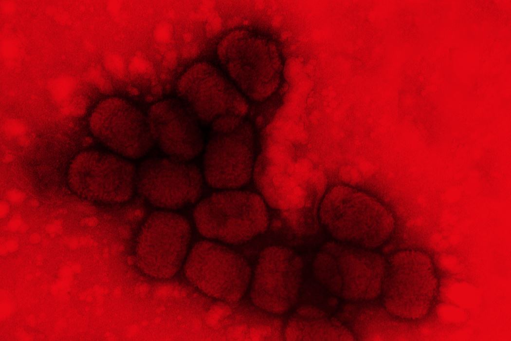
This may explain why RNA viruses generally cause more serious diseases, such as polio, measles, rabies, AIDS, COVID-19, and hemorrhagic fever caused by Ebola. Despite the aggressiveness of RNA viruses, one of the most lethal viruses faced by humankind is DNA-based: the smallpox virus, which killed about 300 million people in the twentieth century alone.
A cluster of copies of the smallpox virus, a disease that killed 300 million people worldwide in the twentieth centuryFred Murphy / CDC
Viruses that cause diseases in humans are by far the most studied, although they are relatively few (around 250 species). In many situations, however, viruses are not harmful and can even help the host survive. Some can be used in ways that benefit humans. In an article published in the journal Science in 2007, a team led by American biologist Marilyn Roossinck, at the time a researcher at the Samuel Roberts Noble Foundation, described a mutually beneficial relationship between a virus, a fungus, and a plant found in soils at temperatures of up to 65 degrees Celsius in Yellowstone National Park, USA. Roossinck and her group found the fungus Curvularia protuberata on the grass Dichanthelium lanuginosum, with which it coexists in harmony. In laboratory tests, the researchers found that the fungus and the plant could only withstand such heat because of a protein in a virus that infected the fungus. The virus made it possible for the hosts to survive, which in turn allowed the fungus to grow in a controlled manner, in a relationship the biologists call mutualism.
In other situations, only one of the hosts benefits. This is the case with the parasitic wasps of the Braconidae and Ichneumonidae families; they lay their eggs inside caterpillars, which are eaten by the larvae when they hatch. The larvae are only able to overcome the caterpillar’s defenses because of a virus found in the wasp’s ovary. When it lays its eggs, it infects the caterpillar with the Polydnavirus, which inhibits its immune system. “As a result of this interaction between the virus and the wasp, the insect has been used as a biological pest control method against caterpillars that attack sugarcane plantations,” says virologist Bergmann Ribeiro, from the University of Brasília (UnB), a specialist in baculoviruses, which attack moth and butterfly caterpillars. For decades, these viruses have been sprayed on soybean crops as a pesticide.
Virologists say that whether they are alive or not, viruses have significantly influenced life on Earth. One reason is the way in which many of them interact with their host cells: either by introducing new genes, or by helping to exchange genes between different species when they jump from one to another. The sequencing of the human genome, for example, revealed that 8.3% of our genes are of viral origin. Another factor that increases the diversity of viruses is that they replicate their genetic material very quickly and with less error control, encouraging the accumulation of mutations that can be transmitted to subsequent generations.
Viruses are a major cause of epidemics. In this century alone, there have already been five: three caused by coronaviruses, one by Zika, and another by Ebola. For this reason, and due to the fact that they are so abundant and diverse, experts have long recommended keeping a watchful eye on viruses. In a survey of viruses that cause diseases in humans published in the journal Philosophical Transactions of the Royal Society of London in 2012, zoologist Mark Woolhouse and his colleagues from the University of Edinburgh, Scotland, issued a warning. “It seems almost inevitable that new human viruses will continue to emerge,” they wrote. “For this reason, an effective global surveillance system for novel viruses is needed.”
Projects 1. Replication and cellular effects of rhinovirus in lymphoid tissues (nº 18/25605-6); Grant Mechanism Regular Research Grant; Principal Investigator Eurico de Arruda Neto (USP); Investment R$182,909.72. 2. Infection of lymphoid tissues by influenza virus (nº 15/25975-0); Grant Mechanism Doctoral (PhD) Fellowship; Beneficiary Ítalo de Araújo Castro; Supervisor Eurico de Arruda Neto (USP); Investment R$234,032.57.
This article may be republished online under the CC-BY-NC-ND Creative Commons license. The Pesquisa FAPESP Digital Content Republishing Policy, specified here, must be followed. In summary, the text must not be edited and the author(s) and source (Pesquisa FAPESP) must be credited. Using the HTML button will ensure that these standards are followed. If reproducing only the text, please consult the Digital Republishing Policy.